hyun-eui-kim-image
September 21, 2016
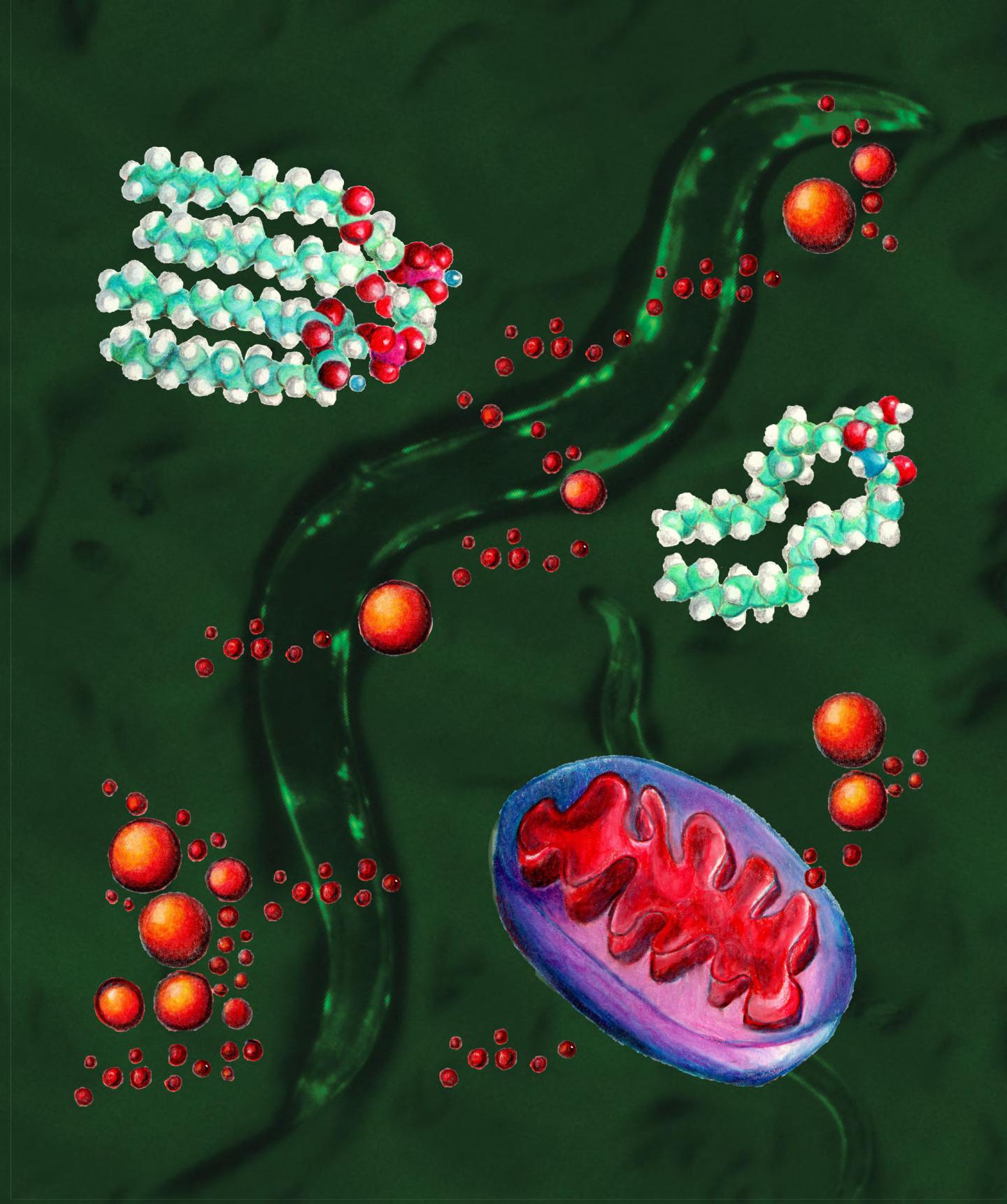
Against a background of nematode worms, this graphic depicts the stressed mitochondria (lower right, blue and red) accumulating lipid droplets (red balls) in the interior or cytosol of the cell. The molecular structure of the two key lipid components in the newly discovered signaling pathway are ceramide (right) and cardiolipin (upper left). The background image shows a picture of the nematode C. elegans with clumps of the Huntington’s aggregates (bright green because they’re tagged with green fluorescent protein) in the body wall muscle cells.
CREDIT
Hyun-eui Kim, UC Berkeley