Class of 2025

University of California, Berkeley
Sponsor: Dr. Alanna SchepartzHarnessing long-range hormone signaling for therapeutic delivery
What do poisonous frog toxins have to do with drug delivery? More than you might think, according to Jane Coffin Childs Fellow Dr. Aurora Alvarez-Buylla.
During her thesis research in Dr. Lauren O’Connell’s lab at Stanford, Alvarez-Buylla identified the first toxin binding protein in poison frogs. This protein, a serine protease inhibitor, or serpin, binds to toxins in the blood, and delivers them to skin glands for bioaccumulation. Interestingly, Alvarez-Buylla found that this frog protein is very similar to mammalian hormone carrier proteins that facilitate the transport of lipid-soluble hormones in the bloodstream and their delivery to target cells.
For the fellowship in Alanna Schepartz’s lab, Dr. Alvarez-Buylla plans to reconstruct the evolutionary trajectory of mammalian hormone carrier proteins. Like frog poison, many therapeutics are toxic when delivered systemically, therefore it is desirable to sequester these therapeutics until delivered to a particular organ. By understanding the range of small molecules that these proteins could possibly bind to, and how their release can be triggered, Alvarez-Buylla’s research will establish a framework for rationally engineering proteins to serve as novel drug delivery agents for many therapeutic molecules.
University of Colorado, Boulder
Sponsor: Dr. Aaron WhiteleyUnlocking the Bacterial Vault: Novel Organelles Involved in RNA Repair
It has been said that RNA is the central molecule in genetic transfer and cellular processes; Dr. Nathan Bullen’s past and planned future research certainly support that sentiment.
During his thesis research in Dr. John Whitney’s lab at McMaster University, Bullen discovered the role of an RNA-modifying enzyme in microbial warfare. Bacteria compete with one another in a microscopic turf war of sorts. One of the ways they combat their foes is by injecting toxins into nearby bacterial cells. Bullen demonstrated that one of these toxins is an enzyme called RhsP2 which works to inhibit protein synthesis or translation in neighboring cells.
As a Fellow in Aaron Whiteley’s lab at the University of Colorado, Dr. Bullen is going on the defensive—asking: how do organisms survive when their RNA is under attack? Intriguingly, the proteins that repair RNA are conserved from bacteria to humans, and Bullen has reason to believe that these systems operate in remarkably similar ways, despite billions of years of evolution. By studying these pathways in bacteria—whose genes are easier to manipulate—his cutting-edge research is shedding light on fundamental processes of RNA metabolism across the tree of life, with far-reaching implications for health, disease, and beyond.
Boston Children's Hospital, Harvard Medical School
Read more
Boston Children's Hospital, Harvard Medical School
Sponsor: Dr. Christopher WalshUncovering oligodendrocyte lineage dynamics in the human brain using somatic mutations
Human brain development is challenging to study for many reasons. Dr. Emre Caglayan’s project as an HHMI-JCC Fellow aims to overcome experimental limitations related to studying brain development and provide unprecedented insight into how a brain develops over the human lifespan.
As a Ph.D. student in Dr. Genevieve Konopka’s lab at UT Southwestern Medical Center, Caglayan investigated human brain evolution. Using advanced genomics technologies, he found that human brains have unique functionalities used for the development and maturation of specialized cells relative to other closely related species.
Dr. Caglayan notes that the absence of non-invasive molecular tools has prevented further exploration of the human brain, and was captivated by the approach of Christopher Walsh’s lab to use somatic mutations as a “barcode” to trace cell lineages. This approach will enable Caglayan’s investigation into brain dynamics so that they can investigate how new mature oligodendrocytes are generated throughout a human lifespan. This research will provide fundamental insight into neurodevelopment and may reveal novel clues about how these processes go awry during neurodegeneration.
Memorial Sloan Kettering Cancer Center
Sponsor: Dr. Junhong ChoiRecording cell states and signaling events to reveal cell fate decisions during pancreatic islet differentiation
Gene transcription, the process of copying DNA into RNA for gene expression, is a complicated process that relies on sequences of DNA known as enhancers to help regulate the process. Enhancers are non-coding stretches of DNA that regulate the expression of a subset of genes.
Dr. Brendan Camellato made crucial insights into enhancer-mediated regulation during his thesis research in Dr. Jef Boeke’s lab at NYU Langone Health. In one of his projects, Camellato investigated transcriptional regulation across various species. In addition to being an impressive technical advance, this approach provided insight into a plausible mechanism for how genetic information is transferred and regulated in yeast and mouse embryonic stem cells.
Now, as a Fellow in Junhong Choi, Ph.D.’s lab at Memorial Sloan Kettering Cancer Center, Dr. Camellato will use enhancers as a tool to record cellular histories during normal development, and in diseased states. Using the recently developed ENhancer-based Genomic Recording of transcriptional Activity in Multiplex (ENGRAM), Camellato will investigate how cell signaling drives stem cell differentiation with single-cell resolution. His research promises to advance these important technologies, as well as provide unprecedented insight into human development.
University of Utah
Sponsor: Dr. Nels EldeEvolution of receptor-ligand selectivity to evade bacterial ligand mimicry
Dr. Lews Caro is fascinated with the molecular arms race that occurs between a host and pathogen and how it shapes their evolution. Caro hypothesizes “that by understanding the molecular mechanisms of evolutionary phenomena, we can actually gain more insight into the evolutionary process itself.”
Caro’s graduate research in Michael Ailion’s lab at the University of Washington uncovered the mechanism of a toxin-antidote system in C. elegans. While such systems are widespread in bacteria and fungi, relatively few examples have been discovered in animals, and those few examples remain poorly characterized.
As a Fellow in Nels Elde’s lab at the University of Utah, Dr. Caro will explore the relationship between a special type of transmembrane protein, called ligand receptors, and the ligand itself. Pathogens secrete toxic ligand imitators which bind to host receptors and hijack normal host signaling. This exerts an evolutionary pressure on the receptor to escape activation by pathogen toxic ligands while retaining responsiveness to host ligands. Caro will use a combination of evolutionary analyses, functional and binding assays, and structural biology approaches to determine how receptors resolve this evolutionary conflict.
Stanford University
Sponsor: Dr. William H. RobinsonDeciphering the Role of EBV-Infected B Cells and Autoimmunity in Post-Infectious Syndromes and Developing a Targeted Elimination Strategy
Illness can interfere with the brain’s ability to function properly, affecting much more than just clear thinking. Dr. Ya’el Courtney’s fellowship research is uncovering how immune responses to illness contribute to neurological dysfunction and may even trigger neurodegenerative processes, revealing new connections between the immune system and the brain.
During her thesis research in Dr. Maria Lehtinen’s lab at Harvard University, Courtney revealed how the choroid plexus, a specialized brain structure that produces cerebrospinal fluid is regulated. She found that secreted factors stimulate the development of specialized neural cells and alter their developmental trajectory. Interestingly, environmental factors such as maternal exposure to certain drugs or immune activation in response to illness also trigger secretion of factors and, eventually, can influence offspring behavior.
Dr. Courtney will examine immune-neurological interplay in Dr. William Robinson’s lab at Stanford University in a different context. There, she’ll examine how autoimmune responses in chronic post-infectious syndromes, such as post-treatment Lyme disease or long COVID, drive neurological manifestations. Courtney’s research has the potential to uncover novel immune-neurological connections and may elucidate novel and therapeutically tractable targets for patients with these long-lasting post-infection syndromes.

Gladstone Institutes
Sponsor: Dr. Alexander MarsonEngineering CAR T cells for Enhanced Cancer Immunotherapy via Protein Interaction Network Analysis
Cancer immunotherapies, such as chimeric antigen receptor (CAR) T cell therapies, have shown great promise against malignancies of the blood but have struggled to effectively treat solid tumors. During his fellowship, Dr. Pascal Devant will focus on understanding how T cells work in an effort to engineer CAR T cell therapies that can better attack solid cancers.
Devant developed his expertise in immunology during his graduate research in Dr. Jonathan Kagan’s lab at Harvard Medical School. There, he focused on caspases, key enzymes that work as the body’s early warning system of invaders. Devant discovered that inflammatory caspases are key enzymes in mammalian innate immunity, providing an alternative activation route to what was previously described. He structurally characterized a caspase complex, providing novel insights into substrate capture and processing.
Now, in Dr. Alex Marson’s lab at Gladstone Institutes, Dr. Devant will generate quantitative protein-protein interaction networks to identify key interactions that regulate T cell function. Devant will leverage this information, using gene editing and preclinical CAR T cell models, to engineer the next generation of CAR T cell therapies for the treatment of solid tumors. In addition to providing fundamental insight into how T cells work, Devant’s work holds great promise in clinical translation for cancer patients.
University of Washington
Sponsor: Dr. Min Yang“Healthy aneuploidy”: Discovering strategies from the placenta to regulate aneuploidy tolerance
Most human cells have two copies of each chromosome, and the loss or gain of entire chromosomes, known as aneuploidy, can often be a characteristic of cancer cells. However, in the placenta, many cells exhibit a high degree of aneuploidy and chromosomal instability. For her fellowship, Dr. Meagan Esbin will study how the cells of the placenta tolerate such high levels of aneuploidy.
During her thesis research, Dr. Esbin studied transcriptional regulation in the joint lab of Drs. Robert Tjian and Xavier Darzacq at UC Berkeley. First, she helped solve the structure of a regulatory hub involved in gene regulation, the human SAGA complex. Then, motivated in part by her passion to improve women’s health and make pregnancy safer, Esbin demonstrated that a human transcription factor (TFEB) plays an essential role in placental cell-cell fusion. This finding reveals new possibilities for rescuing defective cell fusion that can occur in preeclampsia.
In Dr. Min Yang’s lab at the University of Washington, Esbin will take a new angle on understanding placental biology. Using novel cell models, genetic screening, and live imaging she will attempt to define the rules of aneuploidy development and tolerance in the placenta. Her research will provide insight into this life-giving, yet understudied organ. Ultimately, she aims for her research to reveal how we can improve pregnancy outcomes and manage aneuploid cancer cells.

Massachusetts Institute of Technology
Sponsor: Dr. Rebecca LamasonIlluminating the cell envelope architecture and assembly of a tick-borne pathogen
The bacterial cell surface plays a critical role in bacterial physiology and represents a key target for many antibiotics. However, the properties of many bacterial cell surfaces are not well characterized. Dr. Elayne Fivenson’s fellowship project aims to learn more about the cell surface of Rickettsia parkeri, a tick-transmitted bacteria that is a model system for the more pathogenic species Rickettsia rickettsii that causes the deadly Rocky Mountain Spotted Fever (RMSF).
Fivenson developed her expertise in bacterial cell surfaces in Dr. Thomas Bernhardt’s lab at Harvard Medical School. There, Fivenson demonstrated how an inner membrane protein functions to regulate the synthesis of the outer layer of many Gram-negative bacteria. Next, she investigated how the synthesis of the outer membrane and cell wall are coordinated. While it has long been appreciated that the cell wall impacts cell morphology, Fivenson’s results indicate that the outer membrane also contributes to cell shape.
Now in Dr. Rebecca Lamason’s lab at Massachusetts Institute of Technology, Fivenson will study R. parkeri, a model system for the more pathogenic rickettsial species that cause RMSF. She will use structural and proteomic approaches to reveal the composition of the R. parkeri cell envelope. Then, she will use genetic approaches to dissect cell envelope synthesis pathways with the goal of identifying therapeutic targets. As tick range expands due to climate change, RMSF prevalence has increased. Fivenson’s research promises new insights towards the eventual therapeutic inhibition of these deadly bacteria.
University of California, Berkeley
Sponsor: Dr. Eva NogalesFrom Bacteria to Biotechnology: Harnessing Retrons for Precision Medicine
CRISPR-Cas systems have revolutionized how specific genes can be precisely edited. Dr. Grace Hibshman’s fellowship project is focused on how to develop the next generation of genome editors.
During her graduate work in Dr. David Taylor’s lab at the University of Texas, Austin, Hibshman became an expert in the structural and functional characterization of CRISPR-Cas systems.
First, she engineered a more specific genome editing tool with less off-target effects. Then, in a tour de force, Hibshman determined the precise 3D structures of this tool in real-time to understand how it recognizes specific sequences of DNA. Her studies have provided crucial insight into how to improve CRISPR-Cas systems for genome editing.
Now, during her postdoctoral research in Dr. Eva Nogales’s lab at UC Berkeley, Hibshman will study a different set of genetic editing tools focusing on retrons, bacterial elements that can fuse multiple enzymatic activities into a single protein. She’ll use biochemical, structural, and high-throughput mutagenesis approaches to characterize and optimize one such retron. Hibshman’s research may provide us with the latest and greatest genome editor, and she’s betting on its applications in a wide variety of diseases such as cystic fibrosis, Alzheimer’s, and Duchenne muscular dystrophy.

Vanderbilt University Medical Center
Sponsor: Dr. Ivelin GeorgievDefining the targets of the antibody response to natural Oropouche virus infection
Dr. Hannah Itell’s passion for understanding the “ever-evolving virus-host arms race” started during her undergraduate global health studies in India, South Africa, and Brazil. Seeing the impact of viral infection on individuals, families, communities, and entire countries, motivated Itell to dedicate her research career to preventing viral transmission.
In her graduate research in Dr. Julie Overbaugh’s lab at the Fred Hutchinson Cancer Center, Itell focused on identifying human traits that limit HIV severity. She discovered that many virus-fighting genes seen in lab-grown cells function differently than real human immune cells,, demonstrating that common lab models may not reflect what really happens in the body. Next, she identified a gene that regulates a pattern in HIV transmission that was not previously understood.
Now as a Fellow in Dr. Ivelin Georgiev’s lab at Vanderbilt, Itell has switched her focus to the Oropouche virus which is endemic to Brazil.There are currently no vaccines or specific treatments available to prevent or treat infection. Itell will find out how many virus types can be blocked by antibodies and where on the virus the antibodies attach. Itell’s efforts will provide fundamental knowledge about host response to Oropouche virus and directly inform rational vaccine design in the development of antibody therapeutics.
Yale University
Sponsor: Dr. Ruslan MedzhitovUncovering the principles of immune sensing within the central nervous system
Dr. Madeleine Junkins is intrigued by brain-body interactions and how this relationship enables complex behaviors and functions. During her graduate research she investigated thirst suppression in ground squirrels, a hibernating species that can forgo water for months. During her fellowship, Junkins will interrogate collaborative immune-neural responses to illness.
During her thesis research in Dr. Elena Gracheva’s lab at Yale University, Junkins demonstrated that a specialized subset of neurons are activated at low temperatures during hibernation and promotes the release of a hormone that tells the body to hold onto water. Additionally, she found that thirst-sensing neurons in specialized brain areas called the circumventricular organs are functionally suppressed during hibernation. Collectively, Junkins’ research provided a major leap forward for understanding the neural regulation of thirst suppression during hibernation.
As a postdoc in Dr. Ruslan Medzhitov’s lab at Yale, Dr. Junkins will now study how our immune and neural systems collaborate to engage defenses when we’re sick. She will uncover the molecular and cellular components that transform inflammatory signals into neural activity. By manipulating the communication between the immune and neural systems during inflammation, Junkins will provide insight into how these two major body systems interact. This understanding could lead to the identification of novel therapeutic targets for neuroimmune disorders.

University of Washington
Sponsor: Dr. Alexander MeeskeInvestigation of immune systems in multicellular bacteria
Dr. Shoshanna Kahne is interested in bacterial pathways and determining how they change in response to their environment. From Mycobacterium tuberculosis to cyanobacteria, Kahne’s research is creating powerful insights with implications ranging from human disease to environmental impacts.
Kahne’s Ph.D. research in Dr. Heran Darwin’s lab at NYU focused on how proteins are marked for breakdown in the bacteria that causes tuberculosis, Mycobacterium tuberculosis. Kahne discovered a protein that regulates marking an important vitamin-making enzyme for degradation in response to the abundance of the vitamin it helps synthesize. Her findings could help identify new ways to treat this deadly disease.
Now, in Dr. Alex Meeske’s lab at the University of Washington, Kahne will study how cyanobacteria defend themselves against infection by viruses. She is investigating species in the order Nostocales and has identified numerous and diverse potential defense systems in their genomes.
Kahne will test Nostocales hosts against diverse viruses to characterize how they succeed or fail to prevent infection. This work may reveal strategies to harness useful qualities of Nostocales, such as their abilities to fix atmospheric carbon and nitrogen, as well as combat their toxic overgrowths which can poison plants, animals, and humans.
Massachusetts Institute of Technology
Read more
Massachusetts Institute of Technology
Sponsor: Dr. Tyler JacksRole of neoantigen-nonspecific Passenger T cells in cancer-associated immunity and tumor progression
Dr. Dave Klawon is fascinated with the critical, yet disparate roles that our immune system plays in resolving or mediating different diseases. He hypothesizes that comparing productive immune responses during infections with immune responses that fail to resolve in autoimmunity or become dysfunctional in cancer will “reveal precise therapeutic targets capable of tuning the immune response at will”.
Klawon developed his immunology expertise during his graduate research in Dr. Peter Savage’s lab at the University of Chicago. His research there focused on understanding how the immune system recognizes proteins from invaders like viruses or bacteria but knows not to attack the body’s own proteins. Klawon found that a special type of adaptive immune cell, regulatory T cells, selectively suppress self-reactive immune responses during infection to prevent autoimmune disease, thereby providing crucial mechanistic insight into self/non-self discrimination by the immune system.
During his fellowship in Dr. Tyler Jacks’s lab at MIT, Klawon will adjust his research focus to the immune system’s role in cancer. Immunotherapy is a burgeoning and incredibly promising cancer treatment modality, yet many patients fail to respond to current therapeutic options. Klawon notes that tumor-infiltrating T cells are a heterogeneous population that include subsets that either combat tumor growth or suppress the immune response allowing tumors to flourish. His research aims to identify factors driving tumor-enrichment of these disparate populations and reveal novel therapeutic targets that would both promote anti-tumor T cells and inhibit immunosuppressive T cells.
Harvard Medical School
Sponsor: Dr. David GintyOrganisational logic of the spino-parabrachial pathway for light touch
The way our brain senses a soothing touch differs from how it senses a painful one, but how these signals are processed are not well understood. Dr. Anna Lebedeva’s fellowship will leverage novel tools developed during her graduate work to answer this question in conscious, freely behaving mice.
Lebedeva developed her expertise in neuroscience and tool development in Dr. Kenneth Harris’s lab at University College London. There she helped develop Neuropixels 2.0, an implant that can steadily track brain activity in thousands of neurons for over two months. Importantly, this miniaturized implant does not constrain animal behavior, enabling measurements in conscious, freely moving mice and rats. Lebedeva then applied her new tool to uncover why mice make certain behavioral choices versus others that have a greater reward output.
During her postdoctoral research in Dr. David Ginty’s lab at Harvard, Dr. Lebedeva will apply Neuropixels 2.0 to understand how the brain processes signals resulting from touch. The brain region called the parabrachial nucleus (PBN) is thought to be important in this process, yet it is unknown how this information is filtered. Lebedeva will be able to monitor thousands of neurons in the PBN to decipher how touch is communicated in response to different stimuli such as light touch, pinching, and heating or cooling. This research will provide unprecedented detail into neural processing of the light touch pathway.

University of California, San Francisco
Sponsor: Dr. Massimo ScanzianiNeuronal mechanisms of simulations in the brain of sleeping mice
Dr. Amir Levi is interested in understanding the neural mechanisms underlying complex behavior. In his thesis research, Levi used innovative techniques to make keen insights into how we learn. In his fellowship, Levi’s research will provide important insight into how our brains generate “internal models”—mental simulations that allow us to predict and control movements and adapt to changing environments.
During his graduate research in Dr. Eran Stark’s lab at Tel Aviv University, Levi focused on neural mechanisms of learning. Learning is frequently understood as the brain adapting to external cues, yet Levi’s approach involved direct manipulation of neuronal networks and subsequent assessment of behavioral performance. Levi created a visual test where mice choose between two options, and found that they can learn the task in just one session, depending on their past experience and how hard the rule is. He also demonstrated how specific brain circuits can transmit neuronal signals with remarkable accuracy and precision, highlighting the brain’s ability to maintain and even enhance signal integrity during processing. His work suggests that using brain activity to guide learning may help us understand how brain signals lead to behavior.
Now in Dr. Massimo Scanziani’s lab at UC San Francisco, Levi will dissect the neural mechanisms involved in generating internal models. Normally, these internal simulations occur together with actual physical movement, making it challenging to study prediction separately from action. Levi will overcome this limitation by studying mice while they sleep. During sleep, internal models are still generated, but no physical movement occurs. Thus, Levi’s clever approach will enable him to tease apart the neural mechanisms for these distinct functions. His research will help us understand how the brain anticipates events, coordinates movements, and processes experiences during dreams.

University of Utah, Huntsman Cancer Institute
Read more
University of Utah, Huntsman Cancer Institute
Sponsor: Dr. June RoundThe early-life microbiome regulates β-cell function and type-1 diabetes via gamma-aminobutyric acid
Environmental interactions during prenatal development have important implications that often last well into adulthood. Dr. Diego López’s research has shown how infections can alter this developmental trajectory, impacting immune function and influencing the development of asthma. In his fellowship he will investigate how our microbiome impacts developmental trajectories and metabolic outcomes in adulthood.
López’s graduate research began in Dr. Anna Beaudin’s lab at UC Merced focused on how maternal immune activation and inflammation have lasting impacts that continue well into the offspring’s adulthood.
In one project, Lopez found that when a mother’s immune system is activated, it causes an increase in certain early immune cells—and this increase lasts into adulthood. Additionally, he demonstrated that maternal inflammation expands and hyperactivates a specific population of innate immune cells that cause their offspring to have increased risk for developing asthma in adulthood. Collectively, López’ results reveal the long-lasting consequences of maternal immune activation on offspring fitness.
Now in Dr. June Round’s lab at the University of Utah, López will shift his focus to a different type of environmental interaction: our microbiome. This collection of trillions of microorganisms in our gastrointestinal tract plays a key role in the development of numerous diseases, including type-1 diabetes. Recently, the Round lab demonstrated that loss of early-life microbial diversity during a critical developmental window results in lifelong metabolic dysfunction due to reduced beta cell development. López will investigate the molecular crosstalk between specific microbes, immune cells, and pancreatic beta cells. His research will increase our understanding of the development of type-1 diabetes, and may reveal novel therapeutic targets for treating this disease.
The Ragon Institute of MGH, MIT, and Harvard
Sponsor: Dr. Harikesh WongA spatial stochasticity theory resolves how host-protective T cell responses emerge amid regulatory T cell immunosuppression
Dr. Tomer Milo appreciates distilling simplicity out of complex biological systems. During his graduate work, Milo developed elegant theories for a variety of human diseases and collaborated with experimentalists to validate them. In his fellowship he will develop his own experimental expertise and combine it with his theoretical expertise to tease apart immune processing of self vs. foreign antigens.
During Dr. Milo’s thesis research in Dr. Uri Alon’s lab at the Weizmann Institute of Science he “studied design principles of physiological systems to better understand complex human diseases.” His work provided groundbreaking insight into the tumor microenvironment, bipolar disorder, and autoimmune disease. In his work, Milo used mathematical modeling to identify molecular players and cellular interactions critical in a host of biological diseases.
As a postdoc in Dr. Harikesh Wong’s lab at the Ragon Institute and Mass General, Dr. Milo will focus on systems immunology. Milo will investigate how a specific immune cell population, regulatory T cells, prevents autoimmune responses to self antigens while allowing appropriate immune responses against pathogenic non-self antigens. He thinks that the spatial segregation of the lymph node is crucial for this discrimination and will use high-resolution imaging and mouse models to tackle this question. Milo’s research will answer critical and fundamental questions in immune biology and provide insight into immune responses at homeostasis, during infection, and in autoimmune disorders.

Rockefeller University
Sponsor: Dr. Kivanc BirsoyA genetic approach to study metabolite sensing and regulation in organelles
Dr. Toshitaka Nakamura is interested in understanding protein-chemical interactions that mediate how cells sense stress. During his graduate work he found and characterized new compounds that kill cancer cells by triggering a type of cell death called ferroptosis. In his fellowship, he is interested in understanding how cells handle iron and glutathione, a crucial antioxidant and detoxifying agent, to mitigate stress responses.
In Dr. Nakamura’s graduate research in Dr. Marcus Conrad’s lab at Helmholtz Munich, he investigated the role of the protein ferroptosis suppressor protein (FSP1) in halting ferroptosis, a form of cell death that functions by damaging cell membranes. Nakamura discovered molecules that block FSP1, which induces cancer cell death. He showed that these molecules work by moving FSP1 away from cell membranes, inactivating the inherent enzymatic activity that protects them from damage. Then, by studying FSP1 mutations from cancer patients and lab experiments, he found another inhibitor and identified out how both types work. Collectively, his research provided groundbreaking insight into the role of FSP1 in ferroptosis, and revealed how this protein can be therapeutically targeted in cancer treatments.
Now, as a fellow in Dr. Kıvanç Birsoy’s lab at Rockefeller, Nakamura will study how cells sense metabolites in different cellular compartments. To facilitate his studies Nakamura will develop a CRISPR-Cas9-based genetic screening platform that can target specific organelles. Then, he’ll leverage his platform to investigate iron and glutathione sensing in mitochondria. In addition to providing a novel, widely applicable research tool, Nakamura’s studies may provide new insights and identify tractable therapeutic targets in diseases like cancer and neurodegeneration.
Princeton University
Sponsor: Dr. Joshua RabinowitzBiochemical basis and function of lipid spatial localization within the brain
Dr. Vanha Pham hypothesizes that there are many molecular examples of the “Goldilocks principle” in cell biology. During her graduate research she demonstrated how the right amount of formaldehyde mediates functional epigenetic signaling, while too much leads to general toxicity. In her fellowship, Pham will investigate how lipids in the brain are organized to maintain homeostasis and the functional and disease implications that result when they are not.
Pham’s thesis research in Dr. Chris Chang’s lab at UC Berkeley, focused on the role of small molecules and metal ions in helping the body’s cells carry out chemical processes. Pham discovered that formaldehyde can block an enzyme needed to make SAM, a key molecule in one-carbon metabolism. Surprisingly, this didn’t cause widespread changes in gene regulation, but instead affected only specific spots on certain genes.
As she transitions to Dr. Joshua Rabinowitz’s lab at Princeton University, Pham will shift her studies to the interplay between proteins and lipids. Using the brain as a model system, she’ll investigate why lipids are spatially enriched in different layers of the brain using a novel approach that maps lipids and gene activity in specific parts of a tissue in combination with genetic screens.
Additionally, she will investigate what happens when certain lipid patterns are disrupted. Pham anticipates that her findings will reveal important insights on how lipids impact membrane protein function, regulate cell morphology, and modulate physiology and disease.
Washington University in St. Louis
Sponsor: Dr. Rui ZhangStructural mechanisms for higher-order microtubule assembly function in parasites
Dr. Matthew Reynolds is fascinated with the elegant structures of our cytoskeleton – a large network consisting of protein fibers and associated proteins that gives shape and structure to cells. During his thesis research he developed machine-learning based techniques to enable the structural determination of curved and bundled actin structures. In his fellowship, Reynolds will detail specialized cytoskeleton super-assemblies from parasitic cells.
During Reynolds’ thesis research in Dr. Greg Alushin’s lab at Rockefeller University, he made important contributions to processes involved in cryo-EM structure determination. Reynolds developed computational techniques that were crucial in reconstructing bent F-actin segments and bundled F-actin that help shape and move cells.
Now, in Dr. Rui Zhang’s lab at Washington University in St. Louis, Reynolds will apply his structural biology expertise to more complex cellular systems. He will continue to investigate the cytoskeleton and will use a combination of cryo-EM and cryo-electron tomography (cryo-ET) to examine microscopic single-cell organisms. These studies will provide mechanistic insights into the nanoscale protein-protein interactions that drive micron-scale cytoskeleton organization in single-celled parasites. His research will likely push forward technological development in structural determination via cryo-EM and cryo-ET. Reynolds anticipates that his findings will inform parasitic disease models and may reveal novel therapeutic targets.
University of California, Berkeley
Sponsor: Dr. Michael RapeDiscovery of silencing factors of the unfolded protein response in cancer
Dr. Heegwang Roh recalls how the COVID-19 pandemic hit during a pivotal moment of his graduate training. During the lockdown, he devoted considerable effort to reading literature on basic biology and became interested in the unfolded protein response (UPR), a cellular stress response triggered by the accumulation of unfolded or misfolded proteins in a cell.
Roh’s thesis research in Dr. Alice Ting’s lab at Stanford University involved a number of innovative projects covering a broad swath of chemical biology. In one project, he created a better way to tag nearby proteins using an enzyme called laccase, fixing safety problems seen in older methods. This new system works well for studying proteins and viewing cells under powerful microscopes. In another project, Roh turned a harmless version of botulinum toxin into a tool for delivering proteins inside cells.
As he transitions to Dr. Michael Rape’s lab at UC Berkeley, Roh will utilize his expertise in tool development to interrogate the UPR. This response is important for cells to respond to stress stimuli, yet the molecular mechanisms by which the UPR is suppressed after the stress is resolved is unknown. Roh will use genetic screens to identify novel UPR suppressors and develop chemical inhibitors for the UPR suppressors. In addition to uncovering novel UPR biology, Roh’s studies will provide new tools for studying UPR in cancer cells and perhaps reveal lead molecules for cancer drug development.
Washington University in St. Louis
Read more
Washington University in St. Louis
Sponsor: Dr. Ting WangThe functional role of transposable element-derived transcripts in cancer progression
Dr. Wesley Saintilnord is interested in how transposable elements (TEs), DNA sequences that can move from one location in a genome to another, can exploit epigenetic pathways that then lead to their aberrant reactivation in cancer cells to rewire gene expression programs. In his fellowship, Saintilnord will examine how TEs functionally contribute to cancer progression.
Saintilnord developed his expertise in epigenetic mechanisms during his Ph.D. research at the University of Kentucky in Dr. Yvonne Fondufe-Mittendorf’s lab now at the Van Andel Institute. In his first project, Saintilnord showed that cadmium exposure changes how many genes are turned on during sperm development by affecting DNA methylation. In another study, he found that certain cancer-associated variants of a histone protein make DNA wrap more tightly, changing how genes are expressed. Collectively, his research demonstrates how environmental exposure, and oncogenic mutations rewire gene expression through epigenetic pathways.
Now, in Dr. Ting Wang’s lab at Washington University in St. Louis, he will dissect why cancer cells take control of TEs for gene regulation and how TE-generated transcripts drive tumorigenesis. He will develop a high-throughput screen to evaluate tumor-enriched TE transcripts in classical cancer phenotypes. Then, Saintilnord will evaluate which of these transcripts encode functional proteins that modulate cell signaling and chromatin dynamics. Saintilnord’s studies will provide fundamental insights into TE biology in cancer cells and may reveal novel therapeutic strategies to combat TE-mediated oncogenic programs.
University of Washington
Sponsor: Dr. David BakerDecoding the Structural Basis of Immunogenicity
Dr. Ellen Shrock envisions a future where novel therapeutics are not seen as dangerous by the immune system. While studying the immune response to SARS -CoV2 in her graduate work, she recognized that even different individuals responded in the same way to the virus. In her fellowship, Shrock is systematically characterizing immunogenicity, the ability of a substance to provoke an immune response, and training models to predict antibody recognition, with the long-term goal of avoiding such features in protein therapeutics.
During her thesis research in Dr. Stephen Elledge’s lab at Harvard Medical School, Shrock studied antibodies from people who had COVID-19 and found they targeted over 800 parts of the virus. She also showed that some parts of these antibodies are built into our genes and help the immune system recognize viruses quickly. Shrock’s research is a giant step forward in understanding immune recognition, with important implications for viral immunoevasion and the design of immunosilent protein therapeutics.
As a postdoc in Dr. David Baker’s lab at the University of Washington, Shrock is taking a systematic approach to more broadly understand antibody recognition. She will execute a large-scale screen to characterize the antibody response against a diverse array of proteins. Shrock will then characterize the epitopes within these proteins and use her results to train an AI model to predict immunogenicity. In addition to providing fundamental learnings on immune recognition, Shrock’s findings will empower the design of future protein therapeutics that are invisible to our immune systems.
Stanford University
Sponsor: Dr. Michelle MonjeDisrupting neuron-glioma interactions in the thalamus for thalamic pediatric low-grade glioma therapy
Dr. Patrick Steadman is passionate about neuroscience and the interplay between neurons and glial cells (support cells in the brain) in physiology and disease. During his graduate research, he examined the interaction between these cell types in normal memory consolidation. In his fellowship, Steadman will now investigate how this interplay impacts pediatric low-grade gliomas.
Steadman’s thesis research in Dr. Paul Frankland’s lab at the University of Toronto, focused on the importance of a specialized glial cell, myelin-forming oligodendrocytes, in memory consolidation. He showed that oligodendrogenesis and de novo myelination in the cortex are promoted by learning. Importantly, when he prevented learning-induced increases in oligodendrogenesis, this impaired memory consolidation. Steadman’s results emphasize the role of glial cells in fine-tuning neural circuits for memory consolidation and retrieval.
Now in Dr. Michelle Monje’s lab at Stanford University, Dr. Steadman will continue to examine glial-neuronal interactions, but in the pathological context of pediatric low-grade gliomas. Recent work from the Monje lab demonstrated that gliomas increase neuronal excitability which promotes tumor growth and disrupts normal brain function. Steadman will investigate the molecular mechanisms mediating glioma progression, and test targeted therapies’ impacts on glioma progression and brain function. This research will provide new insight into pediatric gliomas while taking into account the cognitive impact of potential treatments on patients – an important consideration since children with this disease are typically quite young.

Harvard University
Sponsor: Dr. Chenghua GuUnderstanding energetic and vascular constraints on neurophysiology, encoding and behavior
The brain is a remarkable organ; it shapes our perceptions, memories, and cognitive functions, yet these functions come at a high energetic cost. During Dr. Shivang Sullere’s graduate research he discovered a novel mechanism for pain relief that provides significant insight into the role of endogenous cholinergic circuit, while serving as a potential alternative to opioids. During his fellowship, he will investigate the metabolic, neurophysiological and behavioral consequences of deficient energy supply to active brain regions.
During his Ph.D. research in Dr. Daniel McGehee’s lab at the University of Chicago, Sullere used neurophysiological approaches to explore cholinergic circuits involved in central pain signaling. He identified that activating certain cholinergic centers in the brain helped reduce pain, even in conditions in which opioids no longer worked. He then identified the receptor mechanisms mediating the analgesic effects of this cholinergic circuit.
As he transitions to Dr. Chengua Gu’slab at Harvard University, Sullere will adjust his focus to neurovascular coupling (NVC): a dynamic process that matches local blood flow to areas with high neural activity. He will use genetic mouse models and optical methods to disrupt NVC and evaluate how NVC impacts brain function at metabolic, neurophysiological and behavioral levels. Sullere’s studies will provide foundational insights into NVC and may reveal strategies for correcting metabolic deficits in diseases like Alzheimer’s, dementia, diabetes, and atherosclerosis.
University of California, San Francisco
Read more
University of California, San Francisco
Sponsor: Dr. Loren FrankDynamic interactions of hippocampal-prefrontal circuits for flexible behavior
Dr. Shih-Yi Tseng is interested in how the brain coordinates the many functions we use to navigate our way through the world. During her Ph.D. research she studied a large population of neurons to illustrate that neural coding enables these functions and is distributed throughout the cortex. In her fellowship, Tseng will now decipher how two areas of the brain work together to enable the flexible behavior required for navigation.
Tseng’s thesis research in Dr. Christopher Harvey’s lab at Harvard University examined the functional organization of the cortex in supporting sensing, planning, and action to navigate towards a goal location in dynamically changing environments. By tracking the activity of 90,000 brain cells in mice, Tseng found that information about tasks and behavior is spread out across the cortex. Her work suggests that this part of the brain possesses a vast capacity to integrate complex features of behavior and surroundings to guide decisions.
In Dr. Loren Frank’s lab at UC San Francisco, Tseng will now investigate the dynamic interactions between the hippocampus (HPC) and prefrontal cortex (PFC) to enable flexible behavior. The HPC is important for learning and memory, whereas the PFC is crucial for decision-making. These regions need to coordinate during tasks such as navigation, yet how such coordination occurs is not known. Tseng will use multi-area electrophysiology, optogenetic manipulations, and computational methods to determine the HPC-PFC interactions needed for flexible behavior, and to evaluate how their coupling enables these regions to perform their individual functions. This research will provide fundamental insights into HPC-PFC coupling and may reveal ways in which this process goes awry in neuropsychiatric disorders.

Stanford University
Sponsor: Dr. Karl DeisserothNeural mechanism of behavioral exhaustion
Focusing on a task can often leave us exhausted despite low physical exertion. Dr. Yu Wang is investigating the source of this type of behavioral exhaustion. Building off her thesis research on neural sensing of peripheral metabolic states, she’s primed to make key insights into the brain’s metabolic deficiencies that may lead to our fatigue.
Wang developed her expertise in the neural integration of metabolic states during her Ph.D. research in Dr. Ardem Patapoutian’s and Dr. Li Ye’s labs at The Scripps Research Institute. Specifically, she was interested in how sensory neurons regulate peripheral metabolism, metabolic processes that occur in tissues and organs outside of the central nervous system, and how these neurons coordinate intracellular energy use to sustain activity. Wang demonstrated that somatosensory neurons enervate adipose tissue and modulate adipocyte function by acting as a break on the sympathetic system. Interestingly, she found that the mechanoreceptor PIEZO2 is highly expressed in these neurons, and is required for their brake-like function. Collectively, her research has provided keen insight into the interplay between neural function and peripheral metabolic states.
As a postdoc in Dr. Karl Deisseroth’s lab at Stanford University, Wang will examine the neural mechanisms of behavioral exhaustion. She hypothesizes that repetitive behaviors deplete local energy resources in specific brain regions, ultimately leading to behavioral exhaustion. Wang will combine different mouse models with repetitive behaviors, and assess metabolic and energetic states using metabolomics and imaging. Wang’s studies will provide novel insight into behavioral fatigue, and may inform on better intervention strategies.
Cincinnati Children's Hospital
Sponsor: Dr. Aaron ZornMultifunctional RNA-binding transcription factors coordinate cell states in development
Human development at the earliest stages is a complicated process with many intrinsic and extrinsic cell signals. For her fellowship, Dr. Bailey Weatherbee will investigate the molecular mechanisms of lineage-defining transcription factors that enable early embryonic development.
During her Ph.D. research in Dr. Magdalena Zernicka-Goetz’s lab at the University of Cambridge, Weatherbee developed a cellular model of the human post-implantation embryo. By combining various types of stem cells made by turning on certain genes, she created cell clusters that mimic important stages of early embryo development. Furthermore, Weatherbee used cell models and embryos to investigate the requirement of specific signaling pathways for different cell types in early development. These studies are a major step forward in modeling the earliest steps in embryonic development and will enable numerous follow-up studies by the broader scientific community.
Now, in Dr. Aaron Zorn’s lab at Cincinnati Children’s Hospital, Weatherbee will investigate the molecular mechanisms of two critical lineage-defining transcription factors (TFs). She hypothesizes that in addition to their canonical DNA-binding activities, binding to RNA is also crucial for their function. Weatherbee will use cell and animal models to evaluate the developmental significance of TF-RNA interactions and identify partner proteins that mediate their function. Since mutations that impact RNA regulation occur in several congenital diseases and cancers, Weatherbee anticipates that her findings will inform on novel therapeutic strategies to treat these conditions.
Class of 2024

Max Planck Institute of Immunobiology and Epigenetics
Read more
Max Planck Institute of Immunobiology and Epigenetics
Sponsor: Dr. Ibrahim CisséDirect Visualization of DNA Methyltransferase Oligomerization and Its Influence on Neuronal Gene Regulation
Epigenetic modifications are changes that affect how our genes are turned on or off,
without changing the underlying DNA sequence. These changes can be influenced
by factors like our environment, diet, and lifestyle. The epigenetic modifications of
DNA and chromatin-associated proteins play a crucial role in regulating cell-type
specific gene expression. Methylation of DNA, for example, is associated with
turning genes off via transcriptional repression. In most cells, DNA methylation
occurs in the context of “CG” dinucleotides. Neurons, however, are also methylated
at “CA” sequences.
Dr. Stephen Abini-Agbomson predicts that the oligomerization, or the
process of small molecules joining together to form a larger structure, of DNA
methyltransferases (DNMT) is important for their appropriate genomic localization
and activity. He will use cutting edge single-molecule approaches to investigate the
role of DNMT oligomerization in gene expression during neuronal development
in Dr. Ibrahim Cissé’s lab at the Max Planck Institute of Immunobiology and
Epigenetics. Mis-regulation of DNA methylation is frequently observed in
neurodevelopmental disorders and many types of cancer. Dr. Abini-Agbomson’s
findings may produce new mechanistic insights to inform future therapeutic
targeting of these diseases.
Abini-Agbomson honed his expertise in epigenetics and chromatin biology as
a graduate student in Dr. Karim-Jean Armache’s lab at the New York University
Grossman School of Medicine. There, he demonstrated that the histones encoded
by giant viruses can form nucleosomes. This was quite surprising as previously it
was thought that only eukaryotes have nucleosomes. Additionally, Abini-Agbomson
showed that the lysine methyltransferase SUV420H1 impacts chromatin dynamics
through both enzymatic and non-enzymatic mechanisms. With this research
background, Abini-Agbomson is poised to make breakthrough discoveries on the
impact of epigenetic protein oligomerization in neurodevelopment.

University of Washington/Institute for Protein Design
Read more
University of Washington/Institute for Protein Design
Sponsor: Dr. David BakerDe novo design of extracellular effector for modulating distinct outputs of cell-surface proteins
Cell surface proteins can drive diametrically opposed phenotypic outcomes when bound by ligands, distinct molecules that attach to other specific molecules. Therefore, efforts to rewire membrane protein signaling, introducing a specific function could improve and change how we develop treatments.
Dr. Green Ahn will engineer novel membrane protein effectors in Dr. David Baker’s lab at the University of Washington. Using artificial intelligence protein design tools, Dr. Ahn will design a library of de novo extracellular effectors against particular ectodomains of cell surface proteins and investigate how those effectors impact downstream function. Ahn’s studies will provide fundamental insight into membrane protein signaling and set the stage for future therapeutic targeting of these pathways.
Ahn’s expertise in targeting membrane proteins stems from her graduate studies in Dr. Carolyn Bertozzi’s lab at Stanford University. There Ahn developed the first cell-type-specific degrader for a membrane protein. Ahn built on that study to discover cellular factors that are required for targeted membrane protein degradation. She also helped develop the first de novo designed proteins that trigger membrane protein degradation in collaboration with Dr. David Baker’s lab. With this experience Dr. Ahn is poised to make future discoveries with implications for our fundamental knowledge of protein membrane biology as well as future therapeutic strategies.

Boston Children's Hospital/Harvard Medical School
Read more
Boston Children's Hospital/Harvard Medical School
Sponsor: Dr. Taekjip HaProtein-DNA interactions in DSB repair across scales
Homologous recombination (HR) is an important pathway for error-free DNA strand break repair. Proper repair of DNA breaks is crucial for preventing cancer, as indicated by the number of frequent genetic mutations in this pathway that lead to breast, ovarian, and other types of cancer. Therefore, a better mechanistic understanding of DNA break repair may open up new avenues for therapeutic targeting of cancer.
Dr. Ibraheem Alshareedah is taking a novel approach to investigate DNA break repair in Dr. Taekjip Ha’s lab at Harvard Medical School. It is known that BRCA2 loads RAD51 onto single-stranded DNA (ssDNA), yet HR still occurs in BRCA2-mutant cancers, suggesting that there is redundancy in this pathway. Dr. Alshareedah hypothesizes that RAD52 nanoclusters in cells recruit RAD51 and load it onto ssDNA, even in the absence of functional BRCA2. To investigate this hypothesis, Alshareedah will examine if RAD51, ssDNA, and other DNA-break repair proteins are recruited to RAD52 nanoclusters in cells. He will then determine which RAD52 protein features are required for cluster formation. By understanding the partial redundancies in the DNA repair pathway, Alshareedah’s research may reveal novel targets for treating BRCA2-mutant cancers.
Alshareedah’s expertise in protein and nucleic acid clusters stems from his Ph.D. research in Dr. Priya Banerjee’s lab at the University at Buffalo. There, Alshareedah focused on the formation and material properties of protein-nucleic acid biomolecular condensates. He developed an in-condensate passive micro rheology assay and showed that condensates behave as an elastic solid on short time scales, but like a viscous liquid on long time scales. Alshareedah also used this method to probe the aging process of condensates, which is thought to be related to protein aggregation in neurodegenerative diseases. Now in his postdoctoral research, Alshareedah will investigate the functional importance of RAD52 condensates in BRCA2-mutant cancers.

Princeton University
Sponsor: Dr. Annegret FalknerHormone-mediated changes in neural computations that drive flexible social behaviors
Social interactions and behaviors are mediated by a hormone-sensitive brain network. For example, female mice are sexually receptive only during a certain phase of their estrous cycle around ovulation. Yet, it is still unclear how hormones modulate this network’s circuit architecture and dynamics to dictate changes in behavior.
Dr. Meenakshi Asokan will examine this missing link in Dr. Annegret Falkner’s lab at Princeton University. Dr. Asokan hypothesizes that sex hormones reorganize neural dynamics and functional coupling in crucial hubs for top-down control of sociability to drive flexible female social choice. Asokan will use a behavioral assay and computational tools to quantify the hormone-dependent changes in social motivation and preference. She will then pinpoint the location of these differences in the brain, address how these neural ensembles alter the coding of social interactions and manipulate these regions to verify their causal role in hormone-dependent changes in social behaviors. These studies will provide fundamental insight into how sex hormones rewire information processing to impact social behaviors. This information will be invaluable in situations where steep changes in hormone levels are linked to depressive symptoms such as during post-partum, perimenopausal, and pre-menstrual stages in women.
Asokan built her expertise in understanding how neural circuits influence perception and behaviors in Dr. Daniel Polley’s lab at Harvard University. During her graduate studies, Asokan focused on neural substrates that underlie changes in perception following noise-induced hearing loss. She localized this change to layer 5 cortico-collicular axons that innervate the amygdala and striatum, which explains the exacerbated sound-triggered anxiety and aversiveness in hearing loss patients. Additionally, through multi-regional extracellular recordings and optical measurements, Asokan discovered cooperative plasticity in inputs to amygdala as mice learn to reappraise neutral stimuli as possible threats. Asokan will now use her expertise in linking brain regions to changes in social behavior to investigate how sex hormones influence these processes during her postdoctoral research.
The Scripps Research Institute
Sponsor: Dr. Benjamin F. CravattIlluminating cryptic functional pockets in the human proteome with electrophilic stereoprobes
Allostery is a fundamental biochemical process in which one site on a protein influences the function of a different site on the same protein, even if they are far apart. Given this relationship, allosteric sites are versatile drug targets as they can activate, inhibit, or even provide a new function to the protein depending on the specific ligand. Yet, therapeutic cooption of allosteric sites remains limited, in part, due to the prevalence of invisible, cryptic allosteric sites that only appear upon ligand binding.
Dr. Divya Bezwada aims to transform our understanding of cryptic allosteric sites in Dr. Benjamin Cravatt’s lab at The Scripps Research Institute. There Dr. Bezwada will use a chemoproteomic approach to investigate the prevalence of cryptic allosteric sites across protein paralogs. Bezwada’s research will provide first principles regarding the evolution of cryptic allosteric sites and develop novel chemical tools with broad relevance for biological understanding and therapeutic applications.
Bezwada provided novel insight into cancer metabolism during her doctoral research in Dr. Ralph DeBerardinis’ lab at UT Southwestern Medical Center. There she found that clear cell renal cell carcinomas (ccRCC) have defects in the electron transport chain which suppresses oxidative phosphorylation. This result is consistent with decades of research into cancer metabolism. Unexpectedly, Bezwada found that ccRCC metastases upregulate oxidative phosphorylation and that this change is functionally important for metastasis. Bezwada’s discovery has crucial and paradigm-shifting implications for cancer patient treatment. Now, Bezwada will leverage chemical biology techniques to make her next important insights into human biology and disease during her postdoctoral research.
California Institute of Technology
Sponsor: Dr. Zhen ChenDefining the role of protein homeostasis in spermatogenesis
Traditionally, structural biology efforts have been limited to studying purified samples in isolation. While we have learned a great deal via these efforts, such approaches unfortunately strip away much of the biological context from the sample of interest.
Dr. Julian Braxton will overcome these limitations by using cryo-electron tomography (cryo-ET) to examine proteostasis, or the process by which cells maintain the proper balance, folding, and function of proteins, within sperm cells in Dr. Zhen Chen’s lab at the California Institute of Technology. Proteostasis plays important yet understudied roles in cellular development processes, as the proteome must be reprogrammed to enable new functions. Braxton will apply cellular cryo-ET to analyze such developmental processes in mammalian sperm, where highly specialized functional compartments are assembled. This research will provide foundational understanding into the posttranslational regulation of sperm maturation and expand the frontier of cryo-ET development and analysis.
Braxton’s expertise in proteostasis stems from his graduate studies in Dr. Daniel Southworth’s lab at the University of California, San Francisco. There, Braxton used the related structural technique cryo-EM to reveal the intricate details of how the autophagy-related adapter UBXD1 regulates the hexameric AAA+ chaperone p97. His findings revealed that UBXD1 separates two adjacent p97 protomers to open the p97 ring, allowing for a new mode of substrate entry and/or exit into the p97 central pore. In a related project, Braxton revealed a novel asymmetric state of the mitochondrial chaperone Hsp60 that enables client refolding. In his postdoctoral work, Braxton will expand his structural biology toolkit to include cryo-ET and use this technique to provide unprecedented insight into the role of nuclear proteasomes in spermatogenesis.

University of Washington
Sponsor: Dr. Jay ShendureDeciphering the dynamic regulation of mitochondrial genomes
Mitochondria are cellular organelles that house their own DNA. There are hundreds to thousands of copies of the mitochondrial genome (mtDNA) in each cell. Often, mtDNA copies are not the same; rather, a fraction of them carries mutations. Moreover, the composition of mtDNA varies drastically across cells and cell types. Mitochondrial diseases manifest when the pathogenic mutations reach a high percentage in a substantial fraction of cells. However, it is still unclear how mtDNA mutations expand and how cell-to-cell variation of mtDNA composition is formed.
Dr. Yi Fu will address these questions in Dr. Jay Shendure’s lab at the University of Washington. Dr. Fu will develop a method to accurately genotype mtDNA at single-cell resolution and employ this method to monitor mtDNA mutations during differentiation. Fu will also combine this approach with CRISPR perturbation to identify factors that impact the mitochondrial mutation burden in various cell types. These experiments will uncover cell type-specific regulation of mitochondrial genome maintenance. Furthermore, Fu’s research may provide insight into novel therapeutic approaches for mtDNA-associated diseases.
Fu’s expertise in mtDNA stems from her graduate studies in Dr. Agnel Sfeir’s lab at New York University and Memorial Sloan Kettering Cancer Center. There Fu discovered that double-strand breaks in mtDNA activate the integrated stress response, highlighting the cellular program to cope with defective mitochondrial genome. Fu also investigated mtDNA deletions and their impact on cellular metabolism. Now, Fu will leverage genomics and single-cell technologies to elucidate the dynamic regulation of mtDNA during her postdoctoral research.

Yale University
Sponsor: Dr. Seth B. HerzonNovel Chemical Tools for Targeted Eradication of DNA Repair Proteins and Application to Chemosensitization
Glioblastoma is one of the deadliest forms of brain cancer. All glioblastomas contain fast-growing and aggressive tumor cells. The current standard of care, temozolomide (TMZ), extends patient’s lives by a median of 7 months; however, this chemotherapy only works for a subset of patients, and many of those patients rapidly acquire resistance to this treatment. Additional, more efficacious treatments are direly needed for glioblastoma patients.
Dr. Jarvis Hill’s postdoctoral research in Dr. Seth Herzon’s lab at Yale University aims to enable the next generation of glioblastoma therapies. The Herzon lab recently identified a novel small molecule, KL-50, that is effective against glioblastomas lacking the O6-methylguanine-DNA-methyltransferase (MGMT). However, this small molecule does not work on MGMT-positive glioblastomas. In this research, Dr. Hill will develop tumor-specific MGMT inhibitors that can be combined with KL-50 to treat patients with MGMT-positive glioblastoma.
Part of Dr. Hill’s interest in brain tumors grew out of his Ph.D. research in Dr. David Crich’s lab at the University of Georgia. As an organic chemist, Hill devised a novel synthesis for trisubstituted hydroxylamines. Recognizing that these are underrepresented functional groups in medicinal chemistry, Hill next evaluated the drug-like properties of molecules where he replaced hydrocarbons, ethers, or amines with a trisubstituted hydroxylamine. In contrast with long-standing expectations, Hill found that these substitutions were stable and generally well tolerated. Then, Hill used the trisubstituted hydroxylamine motif as a key structural unit to develop an epidermal growth factor receptor (EGFR) inhibitor with excellent brain penetration, which may be useful for treating brain metastases driven by aberrant EGFR. Now, Dr. Hill will turn his dual focus on synthetic medicinal chemistry and neuro-oncology towards finding glioblastoma therapeutics during his postdoctoral research.
Stanford University
Sponsor: Dr. Liqun LuoNeural Circuit Mechanisms for Balancing Instinct with Experience
Neural circuits have been honed by evolution to enable animals to instinctively survive and reproduce in the world that surrounds them. Mammals, however, also have a distinct ability to weigh primal instinct against experience, allowing us to learn how to appropriately respond based on our unique knowledge of the dynamic world around us. However, how the mammalian brain balances the innate robustness of neural circuits with the flexibility afforded by learning remains unclear.
Dr. Tom Hindmarsh Sten aims to answer these questions as a JCC-HHMI Fellow in Dr. Liqun Luo’s lab at Stanford University. To investigate how instinctive behaviors can be modified by learning, Dr. Hindmarsh Sten will leverage natural variation in the ability of mice to suppress their innate fears and learn how to hunt live prey. He will delineate an anatomical blueprint of neural circuits that mediate evasion and predation, and pinpoint the plastic nodes impacted by learning. These studies will reveal how neural circuits, which have been refined by eons of evolution, are modulated to meet immediate and novel demands in the present.
As a Ph.D. candidate in Dr. Vanessa Ruta’s lab at Rockefeller University, Hindmarsh Sten investigated neural circuits mediating reproduction in fruit flies. He pioneered a novel virtual reality-based behavioral preparation which revealed that sexual arousal in male flies reconfigures how they see and respond to female flies. Additionally, Hindmarsh Sten examined how male flies coordinate aggression amongst rivals with courtship towards females in competitive environments where more than one male fly is vying for each female’s attention. This study revealed neural populations that allow males to rapidly switch between aggression and courtship. With this background, Hindmarsh Sten is primed to investigate how learning modulates innate instinct in mammals.
Harvard University
Sponsor: Dr. Michael DesaiLearning structure in genotype to phenotype maps
Inferring the genetic basis of quantitative traits is foundational to understanding the biological mechanisms that underlie complex phenotypes such as behavior, homeostasis, and disease. Mapping genotype to phenotype has been transformational for understanding and treating diseases controlled by a single gene, or monogenic. However, understanding complex, highly polygenic phenotypes with currently available approaches can take decades of research from fields of researchers to make progress, if the problem is even solvable with current methodologies.
Dr. Caroline Holmes will transform the process of unraveling polygenic phenotypes in Dr. Michael Desai’s lab at Harvard University. Dr. Holmes will develop new computational approaches and use high-throughput experiments to learn the structure of interactions between genes involved in a particular phenotype. Holmes then will test her predictions of interactions with mutational perturbations. Ultimately, Holmes will develop methods to improve the generalizability of genotype to phenotype maps and test their accuracy on a distinct microbe that was not used to train the system. If successful, Holmes’ methods would rapidly catalyze the process of understanding and rationally perturbing polygenic phenotypes.
Holmes’ longstanding interest in both biology and physics dates back to her studies and research as an undergraduate student at Emory University. Her graduate studies emphasized the physics side as Holmes mainly used theoretical approaches in the labs of Dr. Bialek and Dr. Palmer at Princeton University. However, many of Holmes’ research applications were still biological in nature. For example, Holmes demonstrated that non-24 hour circadian periods can compensate for systematic error that arises as a result of seasonality. Holmes will now develop quantitative experimental systems during her postdoctoral research and combine this with her expertise in theoretical approaches to make inroads into complex polygenic phenotypes.
Memorial Sloan Kettering Cancer Center
Sponsor: Dr. John MaciejowskiMechanisms and consequences of APOBEC3 targeting of extrachromosomal DNA
Extrachromosomal DNAs (ecDNAs) are circular DNA elements that amplify oncogenes and mediate chemotherapy resistance. Despite their importance in cancer, currently no therapies directly target these aberrant molecular structures.
Dr. Amer Hossain will investigate innate immune system recognition of ecDNAs to limit their oncogenic potential in Dr. John Maciejowski’s lab at Memorial Sloan Kettering Cancer Center. Dr. Hossain’s research will provide a fundamental understanding of the recognition and processing of ecDNAs by the immune system. Furthermore, his studies may provide insight into defects in this process that lead to cancer, and into therapeutic strategies to reinforce immune clearance of ecDNAs.
Hossain studied bacteria-phage conflicts as a graduate student in Dr. Luciano Marraffini’s lab at The Rockefeller University. Specifically, he developed a novel functional assay to screen for antiphage defense elements, and discovered a DNA glycosylase that inhibits phage replication. At first glance, this might seem like a distant subject from cancer biology. Yet, Hossain notes in many ways the immune-ecDNA conflict mirrors the host-pathogen conflict in that they both involve recognition and degradation of DNA substrates. Therefore, Hossain will apply his expertise to cancer biology during his postdoctoral research.
Dana-Farber Cancer Institute & Harvard Medical School
Read more
Dana-Farber Cancer Institute & Harvard Medical School
Sponsor: Dr. William Kaelin, Jr.Unveiling Novel Therapeutic Targets in Cancer Through Cell-Surface Proteomic Profiling
Dr. Yanyan Hu’s research focuses on discovering new biomarkers to help diagnose, monitor, and treat cancer. In particular, Dr. Hu hypothesizes that studying the tumor cell surface proteome will reveal an abundance of potential therapeutic and diagnostic targets against cancer.
In Dr. William Kaelin, Jr.’s lab at Dana-Farber Cancer Institute, Hu has devised a proximity labeling method that enables the direct quantification of proteins on the surface of cancer cells. Hu will now use this method to examine two types of cancer: clear cell renal cell carcinoma, and tumors with homologous recombination defects. In addition to revealing novel and fundamental information on cancer cell surface proteomes, Hu’s research has direct implications for future diagnostic and therapeutic approaches.
Hu’s Ph.D. research in Dr. Sheng Ding’s lab at Tsinghua University focused on totipotent stem cell biology. Totipotent stem cells are capable of producing every kind of differentiated cell in both embryonic and extraembryonic tissues. Previously, they had only been generated through IVF or SCNT using germline cells. Hu discovered a cocktail of three small molecules that converted mouse pluripotent stem cells into totipotent stem cells. Now Hu will apply her expertise of stem cell biology to explore similar mechanisms – such as cellular plasticity, self-renewal, and differentiation – to cancer biology during her postdoctoral research.
Harvard Medical School
Sponsor: Dr. Bernardo SabatiniTime to stop: neural mechanisms of action termination
Sometimes less is more. Our ability to stop an action is an important aspect of executive control, and the lack of this ability is linked to neuropsychiatric disorders like Obsessive-Compulsive Disorder and Attention-Deficit/Hyperactivity Disorder. Yet, it remains unclear how we make and execute stop decisions.
Dr. Shijia Liu will investigate the neural mechanisms and pathways underlying voluntary stop decisions in Dr. Bernardo Sabatini’s lab at Harvard Medical School. Dr. Liu will focus her studies on how mice voluntarily stop licking in response to the absence of water, as a specific instantiation of the broader question. Liu has designed a “licking-for-water” task that will enable her to dissect this process temporally and in different contexts. She will identify the modes of action and neural pathways that mediate stop decisions using optogenetics, large-scale neural recording, and real-time decoding approaches. Liu’s research will improve our understanding of voluntary stop decisions, related neuropsychiatric disorders, and computational mechanisms for context-dependent behavioral switching.
Liu’s expertise in neuroscience stems from her Ph.D. research in Dr. Sung Han’s lab at the Salk Institute for Biological Studies. Her graduate studies focused on the neural connection between perceived pain and breathing, and how opioid drugs impact this connection. Liu identified two subpopulations of lateral parabrachial nucleus (PBL) neurons that express the m-opioid receptor and project to pain and breathing centers. By manipulating activity at the cellular and molecular levels, Liu discovered how to decouple morphine administration and respiratory depression, which would prevent opioid overdose deaths. With this expertise in involuntary physiological-behavioral connections, Liu will now focus on voluntary decisions and their impact on behavior during her postdoctoral research.
Broad Institute
Current Sponsor: Dr. Nir HacohenAwarded Sponsor: Dr. Darrell J. Irvine
LIGHTing up tumor-associated tertiary lymphoid structures through cytokine pharmacokinetic engineering
Proper functioning of our immune systems depends on the precise timing of an orchestra of molecular events. One such important event is the release of cytokines, which are signaling molecules, into the extracellular space to mediate intercellular communication. For cytokines to exert appropriate immunomodulatory roles, their bioavailability must be strictly yet dynamically regulated in space and time. However, the mechanisms by which the immune system interprets the timing of cytokine release remain poorly understood.
Dr. Tianyang Mao will investigate the temporal encoding of cytokine signaling in anti-tumor immunity in Dr. Darrell Irvine’s lab at the Massachusetts Institute of Technology. Dr. Mao will use a novel controlled drug release technology which enables programmable control over the duration of cytokine exposure in vivo. This unique approach will allow Mao to make novel insights into how cytokine temporal dynamics shape cancer immunosurveillance. Better understanding of the immunological impact of cytokine release kinetics will guide the development of temporally reprogrammed cytokine therapeutics for cancer treatment.
Mao’s expertise in immunology emerged as a graduate student in Dr. Akiko Iwasaki’s lab at Yale University. There Mao developed an intramuscular prime–intranasal boost vaccine strategy for SARS-CoV-2 termed “prime and spike,” which leverages preexisting immunity generated by primary mRNA-LNP vaccines to elicit mucosal immunity within the respiratory tract using unadjuvanted intranasal spike boosters. In addition, he developed several antiviral strategies that trigger type I interferon-based immune protection against SARS-CoV-2, including a short stem-loop RNA agonist for the innate immune receptor RIG-I and an aminoglycoside antibiotic with unexpected antiviral properties. Collectively, these strategies hold great promise to not only prevent disease, but also viral transmission. Now, Mao will build on this experience, using novel bioengineering techniques in the Irvine Lab, to make new inroads into the importance of timing in immune responses to cytokines.
University of Utah
Sponsor: Dr. Alana WelmRon Tyrosine Kinase deficiency uncovers a critical regulator of anti-tumor T Cell responses
Metastasis, which includes the dissemination of tumor cells from a primary site and subsequent colonization of faraway sites, is the primary cause of cancer deaths. This process requires a failure of our immune system to recognize and destroy metastasizing cancer cells. As such, targeting cancer during the metastasis step will help create therapies for patients with many different types of cancers (breast, prostate, colon, etc.).
Dr. Marija Nadjsombati will investigate the immune response during metastasis in Dr. Alana Welm’s lab at the University of Utah. Dr. Nadjsombati will use mouse models of breast cancer which faithfully recapitulate metastatic propensity. Nadjsombati will develop new cancer models and investigate their transcriptional regulatory networks to decipher the role of T cell regulation in metastasis. These studies will provide novel insights on both T cell regulation and on targeted therapies for cancer immunology.
Nadjsombati built her expertise in immunology as a graduate student in Dr. Jakob von Moltke’s lab at the University of Washington. There she studied a specialized type of epithelial cells, called tuft cells, which initiate immune responses in the small intestine. Nadjsombati discovered that succinate triggers the downstream signaling in tuft cells that initiates a type 2 immune response. Additionally, by comparing different mice strains, and performing genetic crosses, Nadjsombati showed that Pou2af2 isoform expression is a key regulatory mechanism that determines tuft cell frequency. With this strong immunological background, Nadjsombati is poised to make new breakthrough discoveries on the immune regulation of metastasis.
NIH/NICHD
Current Sponsor: Dr. Jeffrey FarrellAwarded Sponsor: Dr. Amy Shyer
Linking Parts to Process: Probing the Cell-Biological Basis for Tissue Patterning in Developing Mesenchyme
Organismal development is an elegant progression from a single cell to billions or trillions of different cells that form our tissues and organs. While much is known about development at the molecular level, important questions remain about how subcellular molecular inputs integrate with “supracellular” physical behaviors of large cell collectives to shape our tissues. Little is known about how subcellular and supracellular dynamics relate among the mesenchymal cell types that give rise to all connective tissues including skin.
Dr. Victor Naturale will make inroads into these questions using a novel vertebrate skin cell platform developed in Dr. Amy Shyer’s and Dr. Alan Rodrigues’ lab at The Rockefeller University. Dr. Naturale expects that understanding how biological organization translates across length scales will provide novel insight into diverse areas including cancer microenvironments and mesenchymal birth defects that lack a single genetic cause.
Naturale developed his interest in developmental biology as a graduate student in Dr. Jessica Feldman’s lab at Stanford University. Working largely at the molecular to cellular scale, Naturale discovered that in C. elegans the polarity scaffold PAR-3 and the transmembrane protein HMR-1/E-cadherin collaboratively build polarity networks at epithelial cell-cell contacts. He demonstrated that HMR-1 also communicates cell polarity at the tissue level. Importantly, Naturale additionally identified a novel symmetry breaking cue arising at the supracellular scale due to emergent cell-cell contact patterns. This research, and the beautiful images within, were highlighted on the journal cover. In his postdoctoral research, Naturale will translate his experience identifying supracellular cues to a novel model system with relevance to cancer and developmental diseases.
University of California, Berkeley
Sponsor: Dr. David SavageRedesigning RNA-guided DNA integration system using protein engineering
CRISPR-Cas systems have revolutionized genetic engineering and led to novel genetic medicines. As powerful as these systems are, they have some disadvantages such as their large size and a lack of orientation bias which limits their therapeutic usage. CRISPR-associated transposons (CASTs) are mobile genetic elements that use CRISPR-Cas systems for RNA-guided transposition. CASTs may represent the next generation of genome editors due to their enhanced features relative to CRISPR-Cas. Yet, CASTs still require further optimization to realize this potential.
Dr. Jung-Un Park will engineer novel forms of CASTs to optimize properties for genome editing in Dr. David Savage’s lab at the University of California, Berkeley. Using structural biology, biochemistry, and protein engineering approaches, Dr. Park will enhance the activity of individual CAST proteins, as well as tune the functional association between different CAST proteins. Ultimately, Park’s research will provide vast insight into genome editing and may result in the next generation of gene editing technologies.
Park’s interest in CAST biology stems from his graduate work in Dr. Elizabeth Kellogg’s lab at Cornell University. There, he solved structures for CAST that informed on both RNA-guided and RNA-independent transposition. Park will leverage his extensive knowledge of CAST structural details to optimize this system for genome editing during his postdoctoral work.
Harvard Medical School
Sponsor: Dr. Rachel WilsonNeural circuit computations underlying memory-guided navigation
In adaptive behavior, we take in information from the world around us and use that information to execute certain actions to interact with the surrounding environment. For example, successful navigation requires us to remember the spatial position of a goal and transform that information into actions that will move us towards that goal. Mechanistically, it is still unclear how neural circuits perform these computations.
Dr. Noah Pettit will approach this question using fruit fly interaction with wind direction in Dr. Rachel Wilson’s lab at Harvard Medical School. Dr. Pettit hypothesizes that specific cell types form a circuit that encodes wind direction, maintains it in memory, and transforms this information to influence body movement. Pettit will use multisensory virtual reality, two-photon imaging, and genetic silencing approaches to investigate this circuit at the cellular and molecular levels. These studies will provide a detailed description of how environmental perception is sensed, stored, and translated into action, thereby providing a general framework for understanding these computations in different systems and organisms.
Pettit generated expertise in the underlying neurobiology of spatial learning in Dr. Christopher Harvey’s lab at Harvard Medical School. During his graduate studies, he examined the role of Fos, a transcription factor implicated in memory and spatial learning. Pettit discovered that Fos-induced neurons are more likely to be place cells – cells that are activated when an animal experiences a certain place in its environment. Additionally, Pettit found that the place code degrades when mice voluntarily disengage from a spatial task, suggesting that the internal state exerts a strong influence on place cell activity. With this experience, Pettit will now transition to fruit flies and understanding how these animals transform and respond to external cues.
Stanford University
Sponsor: Dr. Anne BrunetDecoding Aging Neurogenic Niches: Unraveling Somatic Mutations, Clonal Dynamics, and Functional Decline
Aging is associated with decreased cognitive ability and enhanced risk of developing neurodegenerative diseases such as Parkinson’s and Alzheimer’s. The declining function of neural stem cells (NSCs) is partially responsible for these trends in the aging brain. While much is known about the genetics of late-stage neurodegenerative diseases, relatively little is known about changes that lead to the decline in NSC function.
Dr. Daniel Richard will investigate the accumulation of somatic mutations in NSCs in Dr. Anne Brunet’s lab at Stanford University. He will examine how these mutations change NSC gene expression and neuron production. Additionally, Dr. Richard will explore strategies to genetically manipulate somatic mutations to potentially enhance NSC function. Richard’s studies will provide much-needed insight into fundamental NSC biology during aging and may reveal novel therapeutic strategies for neurodegenerative diseases and cognitive decline.
Richard’s interest in the link between genetic changes and aging emerged from his graduate studies in Dr. Terence Capellini’s lab at Harvard University. There, Richard focused on the genetic regulation of knee development. By comparing functional regulatory regions in human and mouse fetal limbs, Richard discovered mutations associated with an increased risk for osteoarthritis later in life. Now, Richard will shift his focus to aging-related biological changes in NSCs and neurodegenerative diseases during his postdoctoral research.

The University of Texas at Austin
Sponsor: Dr. Jason S. McLellanStructure-based vaccine design targeting mpox antigen A27
The international outbreak of mpox (monkeypox) in 2022 incited global health concerns and underscored the need for an innovative vaccine. However, little is known about potential vaccine targets within the causative orthopoxvirus, mpox virus.
Dr. Emily Rundlet will explore the structure and function of potential mpox vaccine targets in Dr. Jason McLellan’s lab at the University of Texas at Austin. Dr. Rundlet will structurally characterize antigen complexes using cryo-EM and X-ray crystallography, which will enable her to probe their function in the viral lifecycle and design vaccine candidates. In sum, Dr. Rundlet’s work is expected to provide valuable insights into mpox biology and pave the way for future mpox vaccines.
Dr. Rundlet developed her expertise in structural biology in Dr. Scott Blanchard’s lab at Weill Cornell Medicine. During her graduate studies, Dr. Rundlet used cryo-EM and single-molecule FRET assays to make important discoveries about protein translation. With these methods, Dr. Rundlet elucidated how the ribosome initiates movement of tRNAs during protein synthesis and demonstrated that mRNA decoding by ribosomes is kinetically and structurally different in humans and bacteria. Now Dr. Rundlet is using her expertise to uncover the structural secrets of orthopoxviruses to guide vaccine design and prevent future outbreaks.
Harvard University
Sponsor: Dr. Naomi PierceSecretory cell innovation in a symbiotic interaction
Social interactions between distinct species are important at ecological scales yet are mediated at the molecular level by the transfer of biomolecules such as small chemicals and proteins between organisms. Symbiosis is an example of a relationship among species where both species benefit from a social behavior or interaction.
Dr. Trey Scott will examine the symbiotic relationship between butterfly larvae in the Lycaenidae family and ants in Dr. Naomi Pierce’s lab at Harvard University. Lycaenid caterpillars secrete nutritious and psychoactive substances that are ingested by ants. Ants, in return, protect their renewable food source, the caterpillar, during its vulnerable developmental stage. Dr. Scott will determine the molecular, cellular, and evolutionary bases for this example of symbiosis. Scott’s research will provide novel insight into social interactions, broadly speaking, including their evolution.
Scott examined social interactions as a graduate student in Dr. Joan Strassmann’s and Dr. David Queller’s labs at Washington University. Although the above example of symbiosis between ants and Lycaenid butterflies is relatively straightforward, most examples of social interactions contain context-dependent elements of both cooperation and conflict. Using Dictyostelium discoideum amoebae and Paraburkholderia bacteria as a model for social interactions, Scott discovered that the bacteria may benefit or be harmed by the amoebae depending on current environmental conditions – in this case, rainfall. Scott proposed that this flexibility helps the amoebae host survive in harsh soils with variable prey. Furthermore, Scott showed how long-term social interactions influence evolutionary adaptation. With this extensive background in social interactions, Scott is poised to make breakthroughs investigating the evolution of symbiosis between butterflies and ants during his postdoctoral research.

Massachusetts Institute of Technology
Sponsor: Dr. Tyler JacksUnderstanding tissue damage in the pre-neoplasia to neoplasia transition of colorectal cancer
Tissue regeneration, in a normal developmental context, and cancer are both forms of cellular proliferation. However, tissue regeneration is regulated and responsive to the surrounding environment, whereas cancer sheds these restraints. Understanding the commonalities and the differences between tissue regeneration and cancer may provide insight into novel avenues for cancer therapeutics.
Dr. Bing Shui will investigate the role of tissue damage in facilitating the early pre-neoplastic to neoplastic transition in colorectal cancer in Dr. Tyler Jacks’ lab at the Massachusetts Institute of Technology. Dr. Shui will examine how tissue damage cooperates with oncogenic mutations to initiate cancer. He will also compare damaged mutant and wildtype cells to identify vulnerabilities that can be leveraged to selectively destroy precancerous cells. Ultimately, a better understanding of the role of tissue damage in this early precancerous transition may reveal novel prophylactic cancer treatments.
Shui’s interest in the relationship between tissue regeneration and cancer burgeoned in Dr. Kevin Haigis’ lab at Harvard University. During his Ph.D. studies, he examined the role of microRNAs (miRNAs) in colon regeneration and colon cancer. First, Shui demonstrated that miRNAs are required for tissue regeneration and miRNA suppression exacerbated colon damage due to failed regeneration. Next, he examined the role of miRNAs in colon cancer and discovered a novel form of posttranslational regulation mediated by oncogenic K-Ras that governs global miRNA function. Now Shui will use his expertise in tissue damage and regeneration to identify vulnerabilities in colorectal cancer during his postdoctoral research.
The Scripps Research Institute
Sponsor: Dr. Keren LaskerTowards a novel tauopathy therapeutic: harnessing biomolecular condensates for targeted protein degradation.
Tauopathies are diseases such as Alzheimer’s that are characterized by the aggregation of tau protein. Unfortunately, no disease-modifying therapies currently exist for tauopathies, and the impact of these diseases will increase as the global population trends towards an aging demographic.
Dr. Alex Stevens will investigate a novel mode for treating tauopathies in Dr. Keren Lasker’s lab at the Scripps Research Institute. Autophagy-based degradation methods are making progress, yet a hallmark of tauopathies is that these solid tau aggregates resist degradation. To circumvent this issue, Dr. Stevens will engineer biomolecular condensates to clear tau aggregates. Stevens’ research will set the foundation for next generation tauopathy therapies and provide a general framework using biomolecular condensates to modulate pathological events.
Stevens investigated how viruses hijack cellular transport mechanisms during his Ph.D. research in Dr. Samara Reck-Peterson’s lab at the University of California, San Diego. By exploring the conflicts between viruses and the host intracellular transport machinery, Stevens discovered a previously unknown transport mechanism that potentiates the innate immune response. His research provides insight into how cells mount a defense against infecting viruses and highlights the important role of cellular transport in this process. Now, Stevens will attempt to rationally hijack autophagy to enable degradation of aggregated tau.
Oregon Health and Science University
Sponsor: Dr. Michael CohenMechanisms of Type I PARP1 Inhibitors in Regulating Cell Fate Decisions in Cancer
Gladstone Institute
Current Sponsor: Dr. Melanie OttAwarded Sponsor: Dr. Britt Koskella
The molecular biology of Obelisk RNAs
The first century of molecular biology discoveries was enabled by the study of Nature’s original molecular biologists: viruses. Viruses and their simpler cousins, sub-viral RNAs are extremely well adapted to manipulate their host cell. By studying how these agents alter their host, scientists have been able to both understand the mechanisms of diseases as well as derive tools to fight them. Yet there is still much we don’t know about viruses, but even less-so about sub-viral RNAs. Obelisk RNAs are a recently discovered class of widespread sub-viral RNAs with small, structured genomes that seem to bear no resemblance to any known biological entity. The study of Obelisk biology then might reveal molecular mechanisms that have yet to be seen.
Dr. Ivan Zheludev will characterize a novel class of sub-viral RNAs, termed Obelisk RNAs, in Dr. Melanie Ott’s lab at the Gladstone Institute of Virology using a model Obelisk-host system based on a human oral bacterium. Using this system, Zheludev will probe how Obelisk RNA replicates and spreads between cells, the function of the Obelisk-encoded protein Oblin-1, and how Obelisk RNA impacts the host bacterium within complex microbial communities such as the human oral microbiome. Zheludev’s studies will provide foundational knowledge for understanding Obelisk RNAs and provide a general framework for investigating sub-viral RNAs.
Zheludev’s interest in sub-viral RNAs stems from his Ph.D. research in Dr. Andrew Fire’s lab at Stanford University. There, Zheludev created a bioinformatic discovery tool and used it to discover a new class of sub-viral RNAs that he named “Obelisk” RNAs. He demonstrated that Obelisk RNAs are widespread, with examples found on every continent, and that they are diverse, having identified roughly 30,000 distinct Obelisks. Further, they are also found in the microbiomes of between five to fifty percent of assayed human donors. Now in his postdoctoral research, Zheludev will investigate Obelisk molecular biology and their host interactions.
The University of Chicago
Sponsor: Dr. Chuan HeSpatially resolved de novo single-cell translatomics to dissect translational control in cancer
Translation is a key step in gene regulation that dynamically responds to cell stress, signaling, and metabolic alterations. While there are techniques that allow for investigating transcription with single-cell resolution, similar tools for examining translation are lacking.
Dr. Zhuoning Zou will develop a method for analyzing translation in single cells from complex samples in Dr. Chuan He’s lab at the University of Chicago. Dr. Zou’s method will use in situ reverse transcription to develop a spatially resolved single-cell translatome profiling method. This method will enable measuring differences in translation between distinct types of cells in a heterogeneous mixture. For example, Zou will apply her method to patient biopsies and surgical colon cancer samples. In such samples this method will distinguish translation between different individual cancer cells as well other types of cells in the tumor microenvironment. This research will uncover the translation landscape in real human tissues, provide novel insights into translation regulation in the tumor microenvironment, and may reveal potential biomarkers for cancer prognosis and targets for future therapies.
Zou developed sensitive methods for monitoring translation and applied them to rare and heterogenous samples in Dr. Wei Xie’s lab at Tsinghua University. During her graduate studies she helped pilot a method that investigates translation of a single mouse oocyte. Zou then applied that method to human oocytes and early embryos to reveal novel and dynamic translational regulation in early embryogenesis. Now Zou will apply her expertise to examine translational regulation in cancer cells and other cells in the surrounding tumor microenvironment during her postdoctoral research.
Class of 2023
Boston University
Sponsor: Dr. Elliott Hagedorn and Dr. Christopher ChenManipulating vascular endocytosis to direct blood and immune cell migration
The vascular system transports blood and immune cells throughout the body. Yet, how these cells selectively cross the endothelium and enter the appropriate cellular tissues is unclear. Dr. Gwendolyn Beacham will explore the fundamental mechanisms underlying this endothelial transmigration in Dr. Elliott Hagedorn’s and Dr. Christopher Chen’s labs at Boston University. Beacham predicts that endocytosis is important for this process and has identified candidate proteins by investigating blood stem cells. She will use zebrafish as a model system to validate her preliminary findings. Then, Beacham will use this understanding to engineer blood vessels with controllable endothelial transmigration in zebrafish and in human cell culture. This research may help improve the efficiencies of cancer therapies that rely on endothelial transmigration, such as bone marrow transplants and engineered CAR T-cells.
As a Ph.D. student in Dr. Gunther Hollopeter’s lab at Cornell University, Beacham investigated clathrin-mediated endocytosis. In particular, she discovered that endocytosis is inactivated via phosphorylation of the clathrin Adaptor Protein 2. These findings revealed a novel regulatory mechanism for endocytosis and set up Dr. Beacham to explore how endocytosis contributes to endothelial transmigration.
University of California, San Diego
Sponsor: Dr. Elizabeth VillaIn situ structure of WT and PD mutant LRRK2 on cellular membranes
Mutations in LRRK2, a multi-domain kinase and GTPase, is the most frequent cause of familial Parkinson’s disease. However, we currently lack the detailed understanding of LRRK2 function that could lead to therapeutics for Parkinson’s. Dr. Siyu Chen will use cryo-EM and cryo-ET to study LRRK2 and its mutants in biochemical reconstitutions and in cells. Dr. Chen will conduct these experiments in Dr. Elizabeth Villa’s lab at the University of California, San Diego. These experiments will directly visualize the molecular mechanisms of LRRK2 and interacting partners’ function in the cell, and how pathogenic mutations disrupt these processes. Therefore, Dr. Chen’s research may inform on novel therapies for Parkinson’s disease.
As a graduate student in Dr. Yuan He’s lab at Northwestern University, Chen studied DNA double-strand break repair. Specifically, Dr. Chen used Cryo-EM to solve two key intermediate states in the non-homologous end-joining pathway (NHEJ). These structures revealed novel interaction surfaces between NHEJ proteins and allowed Dr. Chen to propose a near complete reaction cycle for NHEJ. Dr. Chen will now apply his cryo-EM expertise to LRRK2 and will use cryo-ET to visualize LRRK2 in cells.
University of Utah
Sponsor: Dr. Nels EldeEvolutionary immunology in Tetrahymena
A wide range of organisms – such as nematodes, sea anemones, and bacteria – possess immune defenses to protect themselves against infectious microbes and viruses. Yet studying the interactions between hosts and infectious microbes remains limited to a handful of species. Dr. Katherine Deets will expand this list by examining the interactions between Tetrahymena thermophila and viruses in Dr. Nels Elde’s lab at the University of Utah. Dr. Deets is currently identifying novel viruses that infect Tetrahymena, and working to understand how Tetrahymena defend themselves against these viruses. This research will unlock a new experimental platform with powerful genetic tools for diversifying studies of the evolution of host-virus interactions.
As a graduate student, Deets investigated host-microbe interactions in the context of the mouse small intestine in Dr. Russell Vance’s lab at the University of California, Berkeley. Dr. Deets discovered a novel mode of antigen presentation that only occurs following inflammasome activation. This finding revealed a new connection between innate and adaptive immunity in the intestine. Dr. Deets will use her experience in host-microbe interactions to expand our understanding of immune defense in diverse.
Duke University
Sponsor: Dr. Stefano Di Talia and Dr. Kenneth PossDissecting the Dynamics of Tissue Patterning During Regeneration
Many animals, including zebrafish, have the ability to regenerate limbs, tails, or fins following amputation. The regeneration process is thought to faithfully reconstruct the appendage, yet it is unknown how spatial and temporal dynamics in gene expression and cell-signaling pathways control regrowth. Dr. Rocky Diegmiller will use quantitative imaging approaches to investigate morphological and patterning dynamics in regrowth of the paired zebrafish pectoral fin. Diegmiller will conduct these studies in Dr. Stefano Di Talia’s and Dr. Kenneth Poss’ labs at Duke University. Diegmiller will explore how gene expression patterns are re-formed following amputation, and throughout regeneration. These studies will reveal insights into the dynamics and robustness of regeneration, and will dissect how multiple signaling pathways are integrated to ensure faithful regeneration. Furthermore, these studies will generate quantitative tools for studying regeneration that can be applied to other systems.
As a graduate student, Diegmiller used mathematical models and imaging to investigate developmental biology in Dr. Stanislav Shvartsman’s lab at Princeton University. Specifically, Dr. Diegmiller used the Drosophila germline cyst as a model system to investigate cell polarity and the emergence of symmetry breaking mechanisms in cell clusters. With his multidisciplinary background in developmental biology, Dr. Diegmiller hopes his research will also yield important connections and distinctions between developmental and regenerative pathways.
Vanderbilt University Medical Center
Sponsor: Dr. Eric SkaarDefining how Clostridioides difficile engages in microbial warfare.
Clostridioides difficile infection (CDI) is the leading cause of hospital-acquired and antibiotic-associated intestinal infections. However, we do not currently have a clear understanding of CDI pathogenesis, which impedes the development of additional therapeutic strategies. Dr. Martin Douglass will investigate how CDI overcomes the human microbiota and immune system in Dr. Eric Skaar’s lab at Vanderbilt University Medical Center. Dr. Douglass will examine how CDI competes for nutrients with the microbiota and immune system. Furthermore, Dr. Douglass will identify which CD genes are required for host colonization and persistence. These studies may provide insight into novel therapeutic targets for treating CDI.
As a graduate student in Dr. M. Stephen Trent’s lab at the University of Georgia, Douglass examined the outer membrane of Gram-negative bacteria. Dr. Douglass discovered novel proteins that are required for the transport of lipids to the outer membrane. These studies provide potential therapeutic targets for novel antibiotics and provide Douglass with a solid foundation for interrogating new targets in CDI.
Johns Hopkins University School of Medicine
Sponsor: Dr. Seth BlackshawUsing single cell transcriptomics to understand homeostatic sleep pressure
Sleep disorders are common and negatively impact our quality of life and biological health. Yet, how the brain encodes the need for restorative sleep is poorly understood. Dr. Leah Elias will investigate the cellular circuits and molecular signals that encode sleep pressure in Dr. Seth Blackshaw’s lab at Johns Hopkins University School of Medicine. Using single nucleus RNA sequencing, Dr. Elias has identified a cluster of neurons that are activated by sleep deprivation. Furthermore, she has identified candidate genes that are differentially regulated in response to sleep deprivation. She will leverage these findings to mechanistically dissect sleep signals in the brain at the cellular and molecular levels. Dr. Elias’ research has important implications for the basic biology of sleep and may reveal novel therapeutic targets for sleep and metabolic disorders.
As a PhD student in Dr. Ishmail Abdus-Saboor‘s lab at the University of Pennsylvania, Dr. Elias studied the neural circuitry controlling social touch. Specifically, she identified a new pathway that connects social touch in the skin to reward circuits in the brain. With this background in neural circuitry, Dr. Elias will now investigate how the need for sleep is encoded in the brain.
Stanford University
Sponsor: Dr. Eric KoolIdentification of Small Molecule Ligands for c-Myc mRNA
c-Myc is a transcription factor and an attractive therapeutic target as it drives the majority of human cancers. However, inhibiting c-Myc at the protein level is difficult, in part due to its intrinsically disordered structure. Dr. Sheng Feng aims to circumvent this problem by inhibiting c-Myc mRNA with small molecules. Dr Feng will use a fragment-based approach using an RNA-biased library that is functionalized to improve affinity for RNA. Dr. Feng will tether fragments that bind to adjacent RNA sites to improve binding affinity and selectivity. These experiments will be conducted in Dr. Eric Kool’s lab at Stanford University. Dr. Feng’s research will explore a new route for inhibiting an important target in oncology and represents a general method for inhibiting other difficult protein targets.
As a graduate student in Dr. Stephen Buchwald’s lab at the Massachusetts Institute of Technology, Feng developed copper hydride-catalyzed bond forming reactions that are highly regio- and stereoselective. Such reactions produce important substructures for pharmaceuticals, agrochemicals, and natural products. Dr. Feng’s background in organic chemistry has prepared her to design and prepare small molecule ligand libraries for targeting c-Myc mRNA.
Massachusetts General Hospital
Sponsor: Dr. Luke ChaoIn situ cryoET analysis of cristae biogenesis and morphology
Mitochondria generate energy needed to power cells and multicellular organisms. Wrinkles in the inner mitochondrial membrane, known as cristae, concentrate molecular motors for energy production. However, it is unclear how the wrinkly cristae are formed. Dr. Michelle Fry will use a clever approach to investigate cristae formation in cells. She will introduce candidate protein/protein complexes into parasitic protist mitochondria. These mitochondria are smooth, making them amenable for testing with proteins are sufficient to generate cristae. Dr. Fry will use advanced electron microscopy techniques to image changes in mitochondrial morphology. Fry will conduct these studies in Dr. Luke Chao’s lab at Massachusetts General Hospital. These experiments will provide fundamental insights into mitochondrial biology and may provide clues for mitochondrial pathological dysfunction.
As a graduate student in Dr. Bil Clemons lab at the California Institute of Technology, Fry used structural biology to study the targeting of membrane proteins to the endoplasmic reticulum. Specifically, Dr. Fry captured several structural conformations of a protein chaperone, Get3. Fry demonstrated how conformational flexibility is important for Get3 to integrate multiple regulatory signals (binding partners, client proteins, nucleotide binding and hydrolysis). Dr Fry is now excited to use cryo-electron tomography to capture the conformational landscape of proteins that regulate mitochondrial cristae formation in cells.
University of California, Berkeley
Sponsor: Dr. Andrew DillinThe role of ribosome stalling in the regulation of longevity
Aging is a complex physiological process coordinated across tissues within an organism. Loss of protein homeostasis is a hallmark of aging, yet it is not understood why dysregulation in protein synthesis occurs, and if this dysregulation drives aging pathologies. Dr. Naomi Genuth will investigate these questions in Dr. Andrew Dillin’s lab at the University of California, Berkeley. Dr. Genuth will use C. elegans to visualize protein synthesis patterns in vivo in different tissues during the aging process. Ultimately, Genuth aims to define the molecules that contribute to dysregulation of protein synthesis and see whether manipulation of these molecules can delay and/or prevent the aging process. Dr. Genuth’s research will improve our understanding of changes in protein synthesis during aging at the molecular, cellular, and organismal levels, and may reveal new therapeutic strategies for aging pathologies.
As a Ph.D. student in Dr. Maria Barna’s lab at Stanford University, Genuth investigated the role of translational regulation in gene expression. Specifically, she developed a quantitative roadmap of how ribosome composition changes during human embryonic stem cell differentiation. Dr. Genuth will now investigate protein synthesis during aging in Dr. Dillin‘s lab.
Massachusetts Institute of Technology
Sponsor: Dr. Fan WangCentral control of the autonomic nervous system in chronic pain and anxiety
Medication for chronic pain often leads to addiction. Dr. Nitsan Goldstein thinks this may be because around one third of people experiencing chronic pain also suffer from anxiety. Additionally, anxiety is a strong predictor of chronic pain development. Dr. Goldstein predicts that targeting pain and pain-induced anxiety together may reduce chronic pain symptoms. She has identified neurons that are anxiolytic and will test their functional relationship with pain-induced anxiety and a chronic pain-like state. Goldstein will conduct her experiments in Dr. Fan Wang’s lab at the Massachusetts Institute of Technology. Dr. Goldstein hopes that investigating both the central and peripheral causes of chronic pain and anxiety will open avenues for more effective pain treatments.
As a graduate student in Dr. J. Nicholas Betley’s lab at the University of Pennsylvania, Goldstein investigated how the brain regulates food intake. Specifically, Dr. Goldstein discovered that the activation of hunger circuits enhances dopamine release, which is critical for motivating humans to seek rewards like food. These studies helped reveal new relationships between neural programs and have prepared Dr. Goldstein to investigate the relationship between chronic pain and anxiety.
Harvard University Medical School
Read more
Harvard University Medical School
Sponsor: Dr. L. Stirling ChurchmanUncovering the regulation and function of nuclear mRNA degradation
mRNA degradation is an important step in gene expression that is traditionally thought to occur in the cytoplasm. However, a recent genome-wide study uncovered a class of genes whose transcripts are predicted to be primarily degraded in the nucleus. Yet, it is unclear how and why these mRNAs undergo nuclear degradation. Dr. Chantal Guegler will use both candidate- and screening-based approaches to determine which pathways are important for nuclear mRNA degradation, and how this process influences cellular physiology. Dr. Guegler will conduct this research in Dr. Stirling Churchman’s lab at Harvard Medical School. This work will reveal the key determinants of nuclear mRNA degradation and how this process contributes to gene expression regulation.
As a graduate student, Guegler studied bacterial toxin-antitoxin (TA) systems and their role in protecting against bacteriophage infection in Dr. Michael Laub’s lab at the Massachusetts Institute of Technology. There, Dr. Guegler demonstrated that the RNase toxin ToxN cleaves phage mRNAs to disrupt the translation and assembly of viral particles. Interestingly, Guegler also demonstrated that T4 phage can combat ToxN using the phage-encoded antitoxin TifA that sequesters RNA-bound ToxN to prevent it from degrading additional phage mRNAs. With her background in RNA degradation in bacterial TA systems, Dr. Guegler will now investigate nuclear mRNA degradation in eukaryotic cells.
Fred Hutchinson Cancer Center
Sponsor: Dr. Sue BigginsElucidate the dynamics of kinetochore assembly by single-molecule imaging
Aneuploidy is a hallmark of cancer development and occurs due to defects in chromosome segregation. The kinetochore, a complex consisting of over 100 different types of proteins, is required for the proper segregation of chromosomes. However, we lack an in depth understanding of the step-by-step assembly process resulting in a functional kinetochore due to the extreme molecular and temporal complexity of this complex. Dr. Changkun Hu will reconstitute kinetochore assembly in vitro and use TIRF microscopy to measure individual kinetochore protein recruitment times in Dr. Sue Biggins’ lab at the Fred Hutch. This approach will allow Dr. Hu to determine rate-limiting steps and key regulating mechanisms in kinetochore assembly and will serve as a blueprint for future studies examining the assembly of other large complexes. Furthermore, this work may reveal novel trouble points in chromosome segregation that lead to aneuploidy in cancer.
As a PhD student in Dr. Nicholas Wallace’s lab at Kansas State University, Dr. Hu’s research focused on the repair of DNA double-strand breaks (DSBs). Dr. Hu demonstrated that beta human papillomavirus type 8 protein E6 (8E6), long known to impair traditional DNA-repair pathways, also promotes DNA repair via a mutagenic DSB repair pathway termed alternative end joining. In this way, 8E6 promotes cancer development by increasing genomic instability. Dr. Hu will now pivot to study genome stability at the chromosome level in Dr. Biggins’ lab.
Harvard University
Sponsor: Dr. Mansi SrivastavaHow to build (and rebuild) an animal
Many animals are capable of whole-body regeneration, enabling the regrowth of missing structures to their original size and shape after major amputation. Most studies investigating this phenomenon have focused on the transcriptional control of differentiation from adult pluripotent stem cells. However, Dr. Allison Kann predicts that an important, yet underappreciated, aspect of regeneration is the role of cell adhesion. Regeneration from stem cells requires free progenitor cells to unite and integrate into multicellular tissues and organs. Dr. Kann will use Hofstenia miamia, a genetically tractable invertebrate model system to investigate the disassembly, formation, and remodeling of cellular junctions during regeneration. Kann will conduct these studies in Dr. Mansi Srivastava’s lab at Harvard University. These studies will reveal new principles of regeneration and identify mechanisms that cells use to converge into multicellular structures.
As a graduate student in Dr. Robert Krauss’ lab at Icahn School of Medicine at Mount Sinai, Kann investigated the activation of muscle stem cells. She identified that cytoskeletal regulation is a key driver of muscle stem cell fate decisions and demonstrated how stem cells transduce injury signals into activation. With her background in adult stem cell biology, Dr. Kann is now ready to investigate how cellular interactions between progenitor cells regulate organismal regeneration.
Broad Institute of MIT and Harvard
Sponsor: Dr. Bo XiaDeciphering the Molecular Mechanisms of Alternative Splicing Regulation via Intronic Transposable Elements
Transposable elements (TEs) play a crucial role in genomic regulation by affecting gene functions, particularly in alternative splicing (AS). Among these, intronic TEs are notably abundant in the human genome, numbering over a million instances. Current research has predominantly fixated on individual TEs near splicing sites, neglecting the vast majority of deep intronic TEs. This oversight hampers our understanding of their collective impact on AS and their relevance to developmental and disease phenotypes. To address this gap, we first start with examining the interaction of TEs within the TBXT gene. TBXT is vital in embryonic development and implicated in tail loss in hominoids and chordoma, a bone cancer where TBXT is aberrantly activated. Exploring these interactions will deepen our knowledge of AS regulation and provide insights into personalized cancer treatment by identifying new genetic markers and therapeutic targets. This research seeks to provide a novel framework to study how the interaction between TEs can affect gene function by modulating pre-mRNA splicing. By uncovering the intricacies of TE-induced AS, we seek to unearth new genetic markers and therapeutic targets, offering novel avenues in disease treatment and prevention.
Dana-Farber Cancer Institute
Sponsor: Dr. Lewis CantleyDetermining the mechanism of sugar sensing in the Mondo pathway
Organisms adapt to scarce and bountiful nutrient environments by employing nutrient signaling pathways. Sugar is a rich source of energy and carbon for organisms, Dr. Jose Orozco will explore sugar-sensing pathways using biochemical and genetic approaches to discover sugar-regulated kinases and their roles in metabolic adaptation. Dr. Orozco will conduct his work in Dr. Lewis Cantley’s lab at Dana-Farber Cancer Institute. These studies may reveal a new therapeutic target to alleviate metabolic maladaptive responses to the chronic overconsumption of sugars and carbohydrates.
As a graduate student in Dr. David Sabatini’s lab at Massachusetts Institute of Technology, Orozco investigated the nutrient-regulated pathway that controls the target of rapamycin complex 1 (mTORC1) kinase. Specifically, Dr. Orozco discovered a new amino acid sensor that integrates S-adenosylmethionine levels, identified a metabolic product of glycolysis that communicates with mTORC1, and discovered new genes in the mTORC1 pathway. Dr. Orozco will continue pursuing his interests in the link between metabolism and signal transduction pathways in his investigations of MondoA.
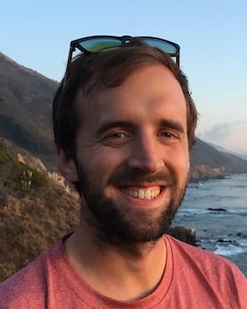
University of California, Berkeley
Sponsor: Dr. Yvette FisherExploring mechanisms for flexible learning in higher-order neural circuits
As we learn new behaviors, we still have to remember old behaviors as well. Thus there is a tension between the flexibility in learning and the stability of maintaining behaviors. Dr. Mark Plitt proposes that neural circuits resolve this tension by using neuromodulation to adaptively switch between stable and labile states. He will investigate these questions in Dr. Yvette Fisher’s lab at the University of California, Berkeley. There, Dr. Plitt will use a fly’s head direction circuit – a neuronal representation of the fly’s orientation in space – to investigate the tradeoffs between flexibility and stability. Dr. Plitt predicts that different neurotransmitters will reinforce learning and maintenance of memory. By developing this powerful model system, Dr. Plitt hopes to uncover physiological and computational principles that govern flexible learning.
As a graduate student in Dr. Lisa Giocomo’s lab at Stanford University, Plitt investigated hippocampal “place” cell remapping – a cellular process that encodes an animal’s memory-guided navigation. Specifically, Dr. Plitt demonstrated that hippocampal remapping patterns are predictably driven by an animal’s prior experience. This expertise in memory establishment will assist Dr. Plitt in investigating the tradeoff between stability and flexibility during adaptive learning.
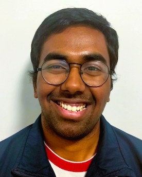
University of California, San Francisco
Read more
University of California, San Francisco
Sponsor: Dr. Massimo ScanzianiTransmission of visual information from the retina to the hippocampus
The hippocampus is a mental GPS that uses visual information to determine relative location. However, the neural pathways that convey visual information to the hippocampus are unknown. Dr. Chinmay Purandare will investigate this information transmission in Dr. Massimo Scanziani’s lab at the University of California, San Francisco. Dr. Purandare will use a novel set of visual cues, developed during his graduate studies, to directly activate hippocampal neurons and determine which visual brain regions are informing the hippocampus. Furthermore, Purandare would probe if the visual information conveyed is different depending on whether the subject is moving versus externally generated visual motion. Dr. Purandare’s research will further our understanding of circuit level connections between visual pathways and the hippocampus.
As a graduate student in Dr. Mayank Mehta’s lab at the University of California, Los Angeles, Purandare explored the minimal set of cues necessary for driving hippocampal responses. He developed novel visual stimuli and found that the hippocampus responds like sensory cortices when presented with these cues. This research led Dr. Purandare to the question of how these visual cues reach the hippocampus, which he will now explore in Dr. Scanziani’s lab.
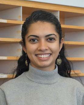
Broad Institute
Sponsor: Dr. Vamsi MoothaPost-transcriptional regulation of mitochondrial biogenesis
Oxidative phosphorylation is a central metabolic pathway that occurs within mitochondria. Decline in oxidative phosphorylation capacity is observed during aging and in many diseases. Dr. Sahana Rao aims to investigate how a tumor suppressor gene also suppresses mitochondrial biogenesis. Dr. Rao will also use a genome-wide screen to identify novel regulators of mitochondrial biogenesis. Rao will conduct these studies in Dr. Vamsi Mootha’s lab at the Broad Institute. Collectively, these studies will provide insight into the regulation of mitochondrial biogenesis. They may also inform on mitochondrial dysregulation in aged or diseased states.
As a graduate student in Dr. Daniel Bachovchin’s lab at Memorial Sloan Kettering Cancer Center, Rao investigated inflammasomes – innate immune sensors that detect pathogenic signals and form large signaling complexes to alert immune cells. Dr. Rao’s studies elucidated molecular mechanisms of the activation of two inflammasome proteins, NLRP1 and CARD8, and established new tools to activate inflammasomes. With her extensive training as a chemical biologist, Rao will now study cellular metabolism and mitochondrial biogenesis in her postdoc.
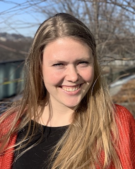
Whitehead Institute for Biomedical Research
Read more
Whitehead Institute for Biomedical Research
Sponsor: Dr. Jonathan Weissman and Dr. Kipp WeiskopfOrigin and function of macrophage heterogeneity in the tumor
Tumor-associated macrophages (TAMs) are the most abundant innate immune cell type in tumors. TAMs can either inhibit or support tumor progression, though it is unclear how their dichotomous functions are regulated. Dr. Alexandra Schnell predicts that the functional heterogeneity of TAMs may be due to distinct lineage origins and cell plasticity. To investigate these hypotheses, Dr. Schnell is developing a myeloid-specific lineage tracing tool to track TAM heterogeneity in tumors, and in response to immunotherapies. Schnell will conduct these experiments in Dr. Jonathan Weissman’s and Dr. Kipp Weiskopf’s labs at the Whitehead Institute. By better understanding TAM heterogeneity, Schnell hopes to enable the development of TAM-targeted cancer immunotherapies that specifically target tumor-promoting macrophages.
During her PhD, Schnell studied the fundamental mechanisms of the immune system in Dr. Vijay Kuchroo’s lab at Harvard Medical School. There, Dr. Schnell performed lineage tracing of immune cells during autoimmune inflammation. Her studies provided a mechanism for how homeostatic intestinal immune cells act as a reservoir for pathogenic inflammation elsewhere in the body. With this background in immunity and lineage tracing, Dr. Schnell will now investigate how the heterogeneity of tumor immune cells can be leveraged to generate new cancer immunotherapies.
University of California, Berkeley
Sponsor: Dr. Jennifer DoudnaPredicting the speed and accuracy of CRISPR-Cas genome editing
CRISPR-Cas enzymes are versatile tools for gene editing and research applications such as transcriptional regulation and imaging. The speed and accuracy of CRISPR-Cas enzymes are crucial, yet how they identify a unique ~ 20-base-pair target within billions of base pairs in the genome is still unclear. Dr. Honglue Shi aims to obtain a more quantitative and predictive understanding of how natural and engineered CRISPR-Cas enzymes rapidly and accurately target specific DNA sequences in Dr. Jennifer Doudna’s lab at the University of California, Berkeley. Shi will use structure-guided biochemistry to develop a kinetic model for CRISPR-Cas9 search speed and accuracy. He will then test the generality of the model on additional CRISPR enzymes and ancestral RNA-guided TnpB enzymes. This research is fundamental to understanding both the evolutionary history of RNA-guided enzymes and the utility of these systems for genome editing. In the future, these results will enable predictions and design of genome editing functions that are not possible or practical today and will greatly accelerate the field as well as the precision and outcomes of next-generation genome editing tools.
As a Ph.D. student in Dr. Hashim Al-Hashimi’s lab at Duke University, Shi focused on the development of biophysical approaches such as NMR spectroscopy to extend the description of nucleic acids from static structures to dynamic ensembles, which results in a deeper and more predictive understanding of how nucleic acids are being recognized by other biomolecules. Having developed this expertise in nucleic acid biophysics and perspectives in dynamic ensembles, Dr. Shi is ready to elucidate the properties that define the best genome editors in Dr. Doudna’s lab.
Duke University
Sponsor: Dr. David SherwoodDetermining how basement membranes stretch and recover to support tissues
Dr. Adam Wei Jian Soh will investigate how the basement membrane (BM), a sheet-like extracellular matrix that encloses tissues, stretches in mechanically-active tissues in Dr. David Sherwood’s lab at Duke University. Dr. Soh will use C elegans ovulation as a novel model system for examining BM stretching and recovery. Soh has performed a localization screen and identified candidate proteins that are likely important for BM dynamics. He will follow up on these findings by determining which proteins are functionally important for the stretching and recovery of BMs. Soh hypothesizes that type IV collagen is critical for stretching tissues as genetic defects in this gene lead to vasculature hemorrhaging and muscle dysfunction. This research may also identify novel genes that are critical for tissue support and are mutated in human disease.
Previously, Dr. Soh investigated the mechanics of motile cilia beating as a PhD student in Dr. Chad Pearson‘s lab at the University of Colorado Anschutz Medical Campus. Specifically, he discovered a novel intracellular mechanism involving the cortical cytoskeleton network that regulates cilia beating synchronization. Through this research Soh developed expertise in imaging techniques and cellular biophysics. This experience has prepared Dr. Soh for his current project dissecting basement membrane dynamics.
University of California, San Francisco
Sponsor: Dr. Loren FrankCortical-hippocampal Neural Dynamics Underlying Model-based Planning
When planning or troubleshooting, we often contemplate possible actions and imagine their outcomes based on prior knowledge. The hippocampus has been implicated in our ability to imagine possible futures, yet it is unclear how future representations are regulated and what functions they subserve. Dr. Xulu Sun will explore the anatomical underpinnings, mechanistic control, and functional significance of hippocampal future representations in Dr. Loren Frank’s lab at the University of California, San Francisco. Dr. Sun will use behavioral tasks and multiregional electrophysiology to explore how the hippocampus interacts with other brain regions to enable future representations and how these representations may support flexible planning. This process is impaired in many neuropsychiatric disorders such as schizophrenia. Thus, Dr. Sun’s research of the underlying neuroscience may reveal new strategies for treating such disorders.
As a PhD student in Dr. Krishna Shenoy‘s lab at Stanford University, Sun investigated dexterous movement control. There she used behavioral tasks and large-scale neural recordings to show how the cortical motor system implements a behavior-organizing map in rhesus monkeys. Dr. Sun will now use her strong foundation in neural computations to explore the neural basis of future representations.
Cornell University
Current Sponsor: Dr. Chris HillAwarded Sponsor: Dr. Elizabeth Kellogg
Mechanism of DNA searching CRISPR-associated transposons
Can we using large language models to talk to our favorite protein sequence and ask it questions? Dr. Jeff Swan thinks we can! He is developing approaches and tools that leverage foundational protein language models to study mechanisms within the AAA+ (ATPases Associated with diverse cellular Activities) protein superfamily in Dr. Chris Hill’s lab at the University of Utah. Combining this with his experience with hardcore wet lab biochemistry, he aims to help bridge the gap between sequence data and protein function.
As a Ph.D. student in Dr. Carrie Partch‘s lab at the University of California at Santa Cruz, Swan investigated the role of the KaiC, an AAA+ protein that effectuates circadian timing. Dr. Swan demonstrated that the ATPase activity in KaiC imparts cooperativity to the transition between autophosphorylation and autodephosphorylation, which is an important feature of the circadian clock. With his expertise in AAA+ proteins, Dr. Swan is primed to uncover the functional hints interwoven in their protein sequence space.
National Institute of Allergy and Infectious Diseases
Read more
National Institute of Allergy and Infectious Diseases
Sponsor: Dr. Michael GriggSexual recombination and virulence in the success of African trypanosomes
In disease-causing organisms, hybridization allows for the transfer of traits such as virulence and drug resistance. Dr. Mabel Tettey will investigate how hybridization impacts African trypanosomiasis outbreaks caused by the parasite Trypanosoma brucei. Dr. Tettey will assess the degree of hybridization occurring in African trypanosome endemic areas, explore the impact of hybridization on virulence, and identify the key molecules involved in this process. She will conduct these experiments in Dr. Michael Grigg’s lab at the National Institute of Allergy and Infectious Diseases. These studies may enable the development of effective disease control strategies against African trypanosomes.
As a graduate student in Dr. Keith Matthews’ lab at the University of Edinburgh, Tettey examined the function of released peptidases in the transmission of African trypanosomes. Specifically, Dr. Tettey identified the genes that dominate quorum sensing signal in African trypanosomes. With her extensive background in trypanosome biology, Dr. Tettey will now examine the role of hybridization in trypanosome virulence.
University of Minnesota
Sponsor: Dr. Anna SelmeckiEffects of regulatory network divergence on drug resistance in Candida
The human pathogen Candida albicans’ genome varies substantially between clinical isolates, yet it is currently unknown how this variation affects infection. Since many genetic variants are located in gene regulatory sequences, Dr. Petra Vande Zande predicts that there is substantial divergence in gene-regulatory networks between different C. albicans isolates that modifies their fitness. Dr. Vande Zande will use gene expression data from different isolates to model gene regulatory networks and identify key differences that impact fitness. Vande Zande will conduct these experiments in Dr. Anna Selmecki’s lab at the University of Minnesota. This research will provide direct insight into genetic differences that impact C. albicans infections. It may also provide clues into other genetically diverse systems with differences in gene-regulatory networks, including human cancers.
As a graduate student in Dr. Patricia Wittkopp’s lab at the University of Michigan, Vande Zande studied gene expression in the context of adaptive evolution. In particular, Dr. Vande Zande discovered that mutations affecting a gene’s expression from a distance are more pleiotropic and more detrimental to fitness than mutations occurring proximally to the gene of interest. With her experience in the evolution of gene expression, Dr. Vande Zande is now interested in understanding divergence in gene-regulatory networks between different clinical isolates of yeast infections.
Harvard University
Sponsor: Dr. Nicholas BellonoMolecular basis of sensory integration in octopus
Cells detect and transform specific external stimuli into precise biochemical functions in a process termed signal transduction. Sensory systems are one example of signal transduction. Dr. Pablo Villar will investigate a unique sensory system: octopus chemotactile receptors that mediate contact-dependent aquatic chemosensation. Dr. Villar will use single-cell sequencing, cryo-EM, and physiology to investigate the molecular logic of receptor expression, complex formation, and physiological function in cephalopods. These experiments will be conducted in Dr. Nicholas Bellono’s lab at Harvard University. Villar’s studies will reveal general principles for the evolutionary fine tuning of signal transduction and help connect adaptations in protein structure with octopus behavior.
As a graduate student in Dr. Ricardo Araneda’s lab at the University of Maryland, Villar examined how neuromodulatory brain regions regulate circuits that process sensory information. Specifically, Dr. Villar showed that the basal forebrain activates shortly after the onset of a sensory stimuli, and in a stimulus-specific manner. With this experience in neuroscience and sensory stimuli, Villar will now examine the signal transduction of stimuli at a molecular level in cephalopods.
University of California, Berkeley
Sponsor: Dr. Eunyong ParkStructural basis of Doa10-mediated protein quality control at the ER
The endoplasmic reticulum (ER) is a critical organelle for maintaining protein quality control in cells; misfolded proteins are targeted for degradation through the ER-associate degradation (ERAD) pathway. Dr. Kevin Wu will study the ER-membrane bound E3 ubiquitin ligase Doa10 in Dr. Eunyong Park’s lab at the University of California, Berkeley. Doa10 is conserved from yeast to humans and identifies and targets many misfolded proteins for degradation. However, it is unclear how Doa10 recognizes a wide range of client proteins. Dr. Wu will use biochemical and structural approaches to reveal how Doa10 recognizes and processes a range of substrates, and how Doa10 cooperates with other quality control factors to maintain protein homeostasis. Protein misfolding and aggregation are associated with aging and diseases such as neurodegeneration. Thus, Wu’s studies may have implications for developing future therapies to improve protein homeostasis in human disease.
As a graduate student in Dr. James Bardwell’s lab at the University of Michigan, Wu investigated chaperone-mediated protein folding. There, he discovered that weak binding between ATP-independent chaperones enable the refolding of client proteins, whereas stronger binding hinders refolding. Dr. Wu’s background in protein refolding set him up for exploring how Doa10 E3 ubiquitin ligase recognizes unfolded protein targets.
Class of 2022

Rockefeller University
Sponsor: Dr. Elaine FuchsInvestigating the roles of spatiotemporal genome architecure in malignancy
Profound alterations in gene expression profiles occur as stem cells from normal tissue transform into cancer stem cells (or tumor-initiating cells) that are able to initiate tumor growth and propagate tumor masses. However, how the gene expression program is rewired during tumorigenesis remains elusive. My research project centers on exploring and identifying key factors that are responsible for driving the divergence of their gene expression profiles. Using in vivo mouse models and genomics and imaging approaches, I am investigating how spatiotemporal genome organization plays a role in oncogenic transcriptional reprogramming during skin squamous cell carcinoma (SCC) development. Skin SCC has emerged as a public health issue due to its increasing incidence and potential for metastasis and recurrence, particularly for the patients placed on immunosuppressive drugs. If successful, my work would further our understanding of the mechanisms underlying oncogenic transcriptional reprogramming and provide new avenues for developing new cancer therapeutics.
University of Texas Southwestern Medical Center
Read more
University of Texas Southwestern Medical Center
Sponsor: Dr. Michael K RosenDecoding the thermodynamics of multi-component biomolecular condensates using high-throughput microfluidics and fluorescence microscopy
Biomolecular Condensates are defined foci found in cells that have selectively concentrated biomolecules. The formation of biomolecular condensates via liquid-liquid phase separation, involving multivalent interactions among biomolecules, has emerged as an important principle for the organization of cellular structure and biochemistry. The molecular underpinnings governing the macroscopic behavior of multi-component condensates, formed by modular protein domains, remain poorly understood. There is limited understanding of how inter-domain binding affinities affect condensate energetics and dynamics, and whether the energetic behaviors can be inferred from known evolutionary relationships between the molecules. Studying these requires quantitative mapping of a phase diagram, which is a two-dimensional array of points for a two-component system and extends to being n-dimensional for n-components. The traditional methods used for mapping phase diagrams require substantial amounts of reagents and are labor-intensive. Hence, I have developed a high-throughput microfluidics-based platform in which hundreds of thousands of pL volume droplets can be made and analyzed as individual reaction chambers in a single experiment, enabling dense sampling of even high-dimensional phase spaces. With this method, I will be mapping phase diagrams and condensate dynamics for systems of increasing complexity. The characterization of the energetics of phase separation with a dataset of unprecedented scale will help in understanding (and eventually predicting) the behaviors of condensates from knowledge of the interactions and co-evolution of individual molecules.
University of California, Berkeley
Sponsor: Dr. Susan MarquseeAn unprecedented atomistic picture of contranslational protein folding
The majority of functions we associate with living thing are made possible thanks to the molecular functions of proteins. Proteins, just like cars and other macroscopic machines, require a specific 3D structure in order to be able to perform their functions and improper protein folding is linked to diseases cut as Alzheimer’s, Parkinson’s, and various cancers. Yet despite decades of research, we still do not understand how proteins fold up into these structures, and what determines whether folding ultimately proceeds correctly—these questions are not addressed by structure-prediction algorithms such as DeepMind’s AlphaFold. It is crucial that we make progress on these issues if we are to rationally design treatments for misfolding diseases, and to predict evolution of organisms, which is often mediated by changes to protein folding and function.
All proteins are made up of one or more chains of amino acids. For some proteins, the physical and chemical interactions between these amino acids are entirely sufficient to drive protein folding into correct native structure. But growing evidence suggests that, for many other proteins, these interactions instead cause the amino acid chain to misfold into non-functional molecular structures. A major goal of my research is to understand how this conundrum is resolved in the complex cellular environment.
One possible resolution to this issue may lie in the fact that, in addition to folding, a protein molecule needs to be synthesized one amino acid at a time by the ribosome. It turns out that many proteins can start folding as they are being synthesized, a process known as co-translational folding which has been shown to significantly increase the odds that certain proteins fold correctly. Indeed, many proteins contain evolutionarily conserved slowdowns in their rate of synthesis at chain lengths corresponding to putative co-translational folding intermediates, indicating it is broadly useful to modulate synthesis rates to give time for co-translational folding. This is akin to how dance (analogous to a chain’s folding) is closely linked to musical rhythm (how quickly amino acids are added)—I may have taken this analogy a bit too far and written a musical piece inspired by it (The Dance of the Nascent Chain).
My research aims to develop a detailed molecular picture of this process, and why it is beneficial to fold co-translationally for many proteins, by combining in vitro and in vivo experimental techniques, physics theory and atomistic simulations. In the future, this interdisciplinary pipeline can also be applied to investigate additional complex processes in the cell including mechanisms of misfolding in disease.
University of Washington
Sponsor: Dr. Michael BruchasEvaluating neuromodulatory networks across brain states
Internal brain states greatly influence our sensations and perception of the external world. Incidents such as stress, hunger, thirst, and pregnancy have all been described as inducing different ‘brain states’ within individuals, altering basic neural properties such as sensory perception, memory, interception, and attention. However, we do not understand how our brains dynamically shape our perceptions and behaviors during state shifts. Here we aimed to create stable brain state models by exposing animals to either chronic social isolation or exercise, two opposing types of behavioral intervention to represent positive and negative experiences. By measuring multi-domain behavioral profiles across the brain during long-term social isolation or exercise, we tested if these biological fingerprints can predict animals’ brain states. To further dissect the neuromodulation changes during brain state shifts, we focused first on the locus coeruleus (LC). LC both receives and sends broad projections throughout the brain. LC cells could then mediate brain-wide changes through its noradrenergic population to control arousal, attention, and sensory perceptions. When simultaneously imaging LCDBH cell body and terminal activities across multiple brain regions, we observed different activity patterns when mice were presented with a diverse array of stimuli. We have also observed dynamic single-cell activities toward different sensory cues, which further confirms the heterogeneity within the LCDBH population. By combing in vivo imaging, circuitry mapping, and biochemical detection, we aim to examine the neuromodulatory signaling dynamics in and out of LC during brain state shifts induced by long-term exercise and social isolation.
University of Michigan
Sponsor: Dr. Kevin WoodInterplay of spatial structure and collective drug resistance across scale
Cells and organisms such as bacteria or cancer cells live in communities and have complex population-level dynamics and emergent behaviors. These behaviors can significantly change their collective properties, including resistance to drugs and other environmental selective pressures. Predicting how cellular communities, rather than individuals, respond to drug exposure could help delay or prevent drug resistance during treatment.
Spatial structure and heterogeneity are important features of such communities – from the cell to the ecosystem scale. In my research, I aim to understand the essential, but poorly understood, relationship between spatial structure and population-level dynamics in bacterial biofilm communities responding to antibiotic selection via theoretical and experimental approaches.
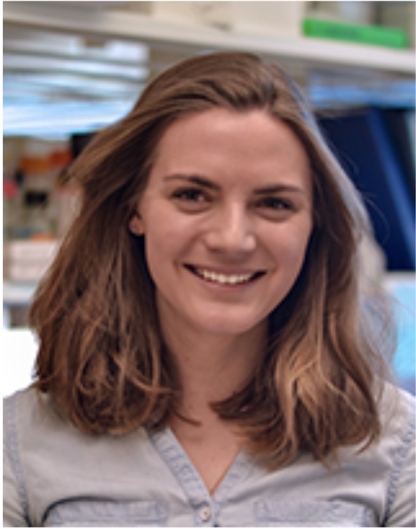
Johns Hopkins University
Sponsor: Dr. Rachel Green and Dr. Bin WuDefining regulatory principles of translation during nutrient shifts
Protein synthesis is metabolically costly; it is therefore critical that cells regulate translation based on nutrient availability. Translation regulation must be flexible to enable cells to adapt to persistent nutrient deprivation, but still recover when nutrients are replenished. Signaling through the kinases GCN2 and mTORC1 can suppress both translation initiation and elongation in response to nutrient depletion, but how each mechanism contributes to global translational control is unclear. Here we investigate translation dynamics upon acute and long-term nutrient depletion, as well as during recovery from nutrient stress. In the immediate response to starvation, GCN2 robustly inhibits initiation to prevent ribosome loading onto transcripts. However, over longer periods of starvation, increased initiation causes translating ribosomes to collide with ribosomes stalled on transcripts. To explore these different temporal regimes, we employ a mass spectrometry-based approach to identify factors that modulate ribosome activity in response to nutrient stress through differential ribosome binding. This work will provide a more integrated understanding of how cells regulate translation and ribosome homeostasis across nutrient environments.
University of California, San Francisco
Sponsor: Dr. Diana LairdOvulation of the fittest
Mammals with ovaries are born with a non-renewing supply of differentiated oocytes ranging from the thousands in mice to the millions in humans. While these high numbers imply a large stockpile, only a comparatively small number of the oocytes present at birth will ever be successfully ovulated and fertilized. To achieve this maturation, an oocyte must first be activated from its quiescent state and then undergo a period of extensive growth in order to accumulate large quantities of biosynthetic materials that are necessary to support the embryo prior to zygotic genome activation. We are currently limited in our understanding of factors that determine whether an oocyte will complete this growth or be fated for elimination/atresia. My research focuses on how both the intrinsic characteristics of oocytes and the extrinsic support provided by the surrounding somatic tissue determine whether an oocyte present at birth will ever be successfully ovulated. My goal is to apply this knowledge to future therapeutics for infertility.
Brigham and Women's Hospital
Sponsor: Dr. Stephen J ElledgeViral manipulation of ubiquitin signaling
Viruses are obligate intracellular pathogens that shape host cell physiology to promote replication and spread. Viral infection is counterbalanced by the host immune response which attempts to detect and eliminate virally infected cells. To reproduce and evade immune detection, viruses modulate expression and post-translational modification of host proteins. The ubiquitin-proteasome system (UPS) mediates targeted degradation of proteins and consists of E1, E2, and E3 enzymes. The modular nature of the ubiquitin ligase pathway has led to co-option by pathogens which alter host protein stability in order to enhance replication and evade immune detection. Understanding how viruses manipulate the host cell proteome to promote infection and how these changes are detected by the immune system is central to the development of anti-viral therapies and vaccines. My postdoctoral research uses genetic and biochemical techniques to discover and dissect host-pathogen interactions with a focus on post translational modifications and their impact on cell signaling.

Harvard University
Sponsor: Dr. Catherine DulacDissecting the neuronal logic of behavioral hierarchy
Animals rely on instinctive behaviors and homeostatic responses, such as parenting, feeding, mating, and sleeping, to ensure individual and species survival. Maximizing survival requires meeting the most pressing needs at the right time, forcing animals to establish behavioral priorities based on a hierarchy of needs. Neurons controlling many of these behaviors are located within the highly interconnected medial preoptic area of the hypothalamus (MPA), making this structure a likely control hub underlying behavioral hierarchy. However, the neural logic of intra-MPA connectivity and how this directs behavioral priorities across physiological states is unknown.
Using the mouse MPA as a model system, I am studying the cell type-specific structural and functional connectivity underlying key competing behavioral and physiological responses. Further, I am determining how animal states, such as virgin or parent, alter neuron function to induce new behavioral priorities. This work will provide the first depiction of a neural basis of the hierarchy of needs and open new avenues for understanding the neural basis of behavior.
Boston Children's Hospital
Sponsor: Dr. Christopher WalshDissecting the impact of non-coding somatic mutations in the human brain
While somatic mutations have been heavily studied in tumors, their prevalence and significance to disease risk in healthy individuals is much less well-understood. The Walsh lab and others revealed that somatic mutation is a widespread phenomenon. Human neurons each contain 100 or more clonal somatic single nucleotide variants (sSNV) at birth, acquired during prenatal development, and gain 15-20 additional sSNVs arising per year. Most somatic variants, including those associated with cancer risk, occur in noncoding regions such as enhancers. Despite being the main source of genetic diversity between cells within an individual, the mechanisms by which noncoding somatic mutations form as well as their functional impact are not well understood. My research will focus on developing new strategies to detect rare noncoding somatic variants as well as dissect their epigenomic impact across different cell types in the human brain. This will help illuminate how much this source of variation contributes to cancer risk and brain disease.

Dana-Farber Cancer Institute
Sponsor: Dr. David PellmanDamage control in response to unreplicated DNA in mitosis
Common fragile sites (CFSs) are hotspots for genomic rearrangements in cancers, but why and how these rearrangements occur is poorly understood. CFSs are characteristically difficult to replicate and often persist as under-replicated DNA into mitosis. Previous work from my host laboratories showed that stalled DNA replication forks are disassembled upon exposure to the mitotic kinase Cyclin B-CDK1. Unloading of the replisome leads to the formation of DNA breaks at the stalled forks followed by break-end ligation events. Coordinated DNA repair events at converging stalled forks in mitosis could lead to the formation of deletions and sister chromatid exchanges, both of which are signatures of CFS expression.
Recent studies have shown that CIP2A is a mitosis-specific repair factor that localizes to sites of DNA damage and replication stress. My aim is to investigate the role of CIP2A in cellular responses to unreplicated DNA in mitosis and how these processes contribute to genomic instability at CFSs. I will use the Xenopus egg extract system to uncover the biochemical mechanism of CIP2A function and conduct cell-based studies to observe the effects of CIP2A on genome stability.
University of California, Berkeley
Sponsor: Dr. Christopher ChangMain-group platforms for deciphering and drugging metionine
It has long been a focus for the identification of protein drivers and for the development of corresponding cancer therapeutics. Traditionally, drug discovery efforts mainly rely on “druggable” proteins, which possess easily identifiable binding pockets or catalytic active sites. However, over 85% of the proteome is still considered “undruggable”, posing additional challenges for further development. Recently, Activity-Based Protein Profiling, has arisen to spotlight the undruggable proteome via covalent linkage of reactivity-based chemical probes and “ligandable hotspots” in the proteome. In the proposed research, I aim to develop new chemoproteomic platforms based on inexpensive and biocompatible main-group molecules for chemoselective methionine and methionine sulfoxide bioconjugation, to further promote cancer drug discovery. This contribution will be significant because it can develop a ligandability map against undruggable proteome, serve as efficient tools in cancer cell early-stage diagnosis, and further provide a handle to decipher and drug methionine redox regulation in cancer cells, thus yielding novel therapeutics.

Harvard University Medical School
Read more
Harvard University Medical School
Sponsor: Dr. Jun R HuhIn Vivo tracking of G protein-coupled receptors sensing for gut metabolites
The human gastrointestinal tract harbors trillions of microbes that have been coevolving with humans for a long time. Growing evidence suggested that the gut microbiota produces a myriad of metabolites, and some of these small molecules possess bioactivity that can shape host development and fitness, such as modulating gut immune cells and promoting brain development. G protein-coupled receptors (GPCRs) represent the largest class of membrane receptors that relay extracellular cues into a cellular response. Many of these GPCRs including orphan GPCRs may evolutionally be designed for communicating with microbes through microbial metabolites. My research seeks to develop a genetic tool and platform that can characterize ligand-activated GPCRs in vivo and uncover GPCRs that sense microbial metabolites. This work potentially sheds light to understand the underlying mechanisms of host-microbiota interaction.
Broad Institute
Sponsor: Dr. Stuart SchreiberA DNA-encoded library strategy for molecular glue discovery
There is an urgent need for cancer therapeutics with improved target specificity and novel mechanisms of action. Molecular glues combine the demonstrated capabilities of small molecules as potent drugs with the power of chemically induced proximity. In enabling new protein-protein associations, molecular glues can address many shortcomings of current small molecule cancer therapeutics, which are often limited to protein targets presenting a clear binding pocket. Furthermore, since protein-protein interactions are widely known to facilitate a range of fundamental cellular activities, chemical compounds which intercede on these pathways can provide access to novel mechanisms of action and enhanced target specificity. The wide-ranging therapeutic potential of molecular glue has already been recognized; however, to date the discovery of these compounds has been limited to serendipity or the synthesis of bifunctional molecules. The systematic and generalizable path to molecular glue discovery has not yet been established. In my research, I will leverage the recording and reporting power of DNA encoded libraries to deliver a new path to molecular glue discovery

Fred Hutchinson Cancer Center
Sponsor: Dr. Sue BigginsDetermining regulation and function of a major microtubule binding pathway
Each time a cell divides, it must ensure equal segregation of chromosomes. Error in this process causes either loss or gain of chromosomes, resulting in aneuploidy, a hallmark of cancer and other diseases. Chromosome segregation is mediated by a megadalton protein complex called kinetochore that assembles at the centromere of each chromosome and serves as the physical linker between chromosomes and the microtubules. Early in mitosis, microtubule-kinetochore attachments are stabilized by tension that distinguishes proper attachment from the improper ones. However, during anaphase, kinetochore-microtubule attachments become vulnerable as tension drops when the chromosomes separate, and the microtubules start shortening. It is major question how kinetochores remain attached to microtubules under low tension. There are two competitive pathways that recruit the major microtubule binding protein, Ndc80c to the kinetochore- Mis12c and CENP-TCnn1 pathway. The CENP-TCnn1 pathway gets enriched at the kinetochore during anaphase, making it a potential pathway that could stabilize low tension attachments. I hypothesize that the CENP-TCnn1 pathway is key to understanding how kinetochores adapt to low tension during anaphase. My goals are to uncover the underlying regulatory mechanism facilitating upregulation of this pathway at the kinetochore during anaphase and how it contributes to kinetochore-microtubule attachments under low tension.
University of Washington
Sponsor: Dr. Alex MeeskeMechanisms of Immune Memory Generation in a Native RNA Targeting CRISPR System
CRISPR systems are the adaptive immune systems of bacteria that are crucial for defense against bacteriophage infection. Immune memory is stored as short DNA sequences in the CRISPR array, called “spacers”, and upon transcription and processing these associate with Cas nucleases to search for matching viral targets and initiate nucleic acid cleavage. Type VI CRISPR systems are unique in that they recognize RNA, and target recognition leads the nuclease Cas13 to indiscriminately cleave cellular RNA. While the targeting steps of this CRISPR type are well-understood, it is still unknown how new spacers are acquired, especially since most type VI CRISPR operons lack the known acquisition machinery. Here, we probe the mechanisms of type VI CRISPR immune memory generation using Listeria seeligeri, a genetically tractable host that endogenously encodes type VI CRISPRs. We show that type VI CRISPR can use the adaptation genes from other CRISPR systems in the genome to integrate new memories into the type VI array, both in vivo and in vitro. In addition, we find no clear bias for acquisition of functional, RNA targeting spacers during growth or infection; however, we do observe some bias for acquisition from highly transcribed regions. In the future, we aim to identify additional factors required for acquisition of new spacers in the type VI CRISPR locus and determine the origin of newly acquired spacers.

University of Washington
Sponsor: Dr. Jakob MoltkeTissue Specific Sentinels of Type 2 Immunity
The innate immune system is paramount in recognizing foreign or mutated material and initiating proper immune responses to combat them. Recognition of allergens and parasitic worms (helminths) elicit a socalled “type 2” immune response focused on expulsion of stimuli and tissue repair. Type 2 immune responses impact the prognosis of many cancers and the success of immunotherapies, but how these responses are established remains poorly understood. Group 2 innate lymphoid cells (ILC2s) initiate and propagate type 2 immune responses, but do not sense immune agonists directly. The origin and regulation of host-derived signals leading to ILC2 activation is therefore an area of immense interest. Recent work identified a specialized population of epithelial tuft cells responsible for sensing helminths and activating ILC2s by secreting interleukin(IL)-25 and cysteinyl leukotrienes in the small intestine. Airway ILC2s are similarly important for type 2 immune responses in the lung, but tuft cells are dispensable in this context. My proposal seeks to identify the signals that activate intestinal tuft cells and a novel cell subset responsible for airway ILC2 activation. Examining the initiation of type 2 immunity in multiple organs will uncover both convergent and divergent mechanisms by which type 2 responses can be further manipulated.

Columbia University
Sponsor: Dr. Rene HenNeuronal and immunological dissection of antidepresant responsiveness
Major depressive disorder (MDD) is a psychiatric disorder with a lifetime prevalence of ~15% and is the leading cause of disability worldwide1. The societal burden of MDD is immense, causing profound personal suffering and economic loss, which has recently been intensified by the Covid-19 pandemic2. The most effective treatments for MDD, a class of antidepressants called the selective serotonin reuptake inhibitors (SSRIs), are successful in achieving remission, but only in ~40% of patients3. Despite being in use for over 50 years, it remains unknown how SSRIs modulate neural circuit function in patients that achieve remission and where these mechanisms are disrupted in those that do not. Thus, a fundamental question remains: What are cellular and molecular mechanisms that mediate antidepressant response and resistance? Defining the answers to this question could provide fundamental insights into the pathophysiology of MDD and uncover novel substrates for future precision medicine approaches.
University of California, San Francisco
Sponsor: Dr. Edward ChangUncovering the functional architecture of the human speech cortex
Speech is a defining characteristic of human cognition. It provides humans with the flexibility to convey an unlimited range of thoughts and feelings using a limited number of basic elements. Over the past decade, intracranial electrocorticography (ECOG) recordings in patients have provided invaluable insights into the neural mechanisms underlying speech perception and production. While significant progress has been made, basic questions still remain regarding the functional architecture of the neuronal circuits involved. Particularly, we do not know how the brain assembles phonemes into words, and words into meaningful goal-directed utterances. Such phonemic-to-semantic transformation relies on real-time interactions between the speech cortex and distributed memory networks that encode, store, and retrieve our lexical and semantic knowledge quickly and efficiently. The hippocampus, as a critical node in this declarative memory system, is believed to play a key role in coordinating such processes in real time.
My research seeks to elucidate the cortical-hippocampal interaction during speech perception and production, and more broadly, to unravel the interface between speech representations and long-term memory. To accomplish this, I combine ECOG recordings with 7T fMRI to measure neuronal activity simultaneously from the hippocampus and speech cortex during perception and production of speech.
Harvard University
Sponsor: Dr. Xiaowei ZhuangReconstruct cell trajectories and communications in brain development
During mammalian development, coordinated cell differentiation and migration convert a simple neural tube into a brain with more than a hundred anatomical regions and probably more than a thousand cell types. How do these cell types emerge? How do cells migrate to their destined locations? How do cells communicate with each other? These are some fundamental problems in brain development.
As a postdoctoral fellow in Xiaowei Zhuang’s lab at Harvard, I develop new methods to systematically study these problems in mouse brain development. I develop new computational methods to connect cells from MERFISH spatial transcriptomics measurements into trajectories and determine cell-cell communication pathways activated in each cell. The reconstructed trajectories will allow me to comprehensively map the differentiation, maturation, and migration of individual cells. I will identify which cell-cell communication pathways are functionally crucial for generating each cell type. Then I will develop high throughput imaging-based screen methods to validate the discoveries.
Columbia University
Sponsor: Dr. Mohammad Al QuraishiLearning the modular grammar of phase separaton in signaling networks
Cells efficiently convert environmental information into specific functional responses through cascades of biochemical reactions and biomolecular interactions. High fidelity signal transduction requires spatiotemporal regulation of these molecular events. This can be accomplished through phase separation. Many signaling condensates dynamically assemble through multivalent protein–protein interactions mediated by modular interaction domains. How the molecular factors that drive phase separation enable coordinated and precise flow of information among myriad signaling pathways remains a mystery. To answer such questions that encompass molecular- and systems-level phenomena, my research focuses on developing integrative data- and physics-based modeling frameworks using the tools of machine learning and statistical mechanics. Using these approaches, I aim to decipher the modular grammar of signaling proteins that governs phase separation and, more broadly, the biophysical principles that underlie cell homeostasis.

University of Colorado, Boulder
Sponsor: Dr. Thomas CechDisseing the impact of non-coding somatic mutations in the human brain
Histone methyltransferase PRC2 (Polycomb Repressive Complex 2) silences genes via successively attaching three methyl groups to lysine 27 of histone H3 (H3K27me3). Several research groups including ours demonstrated that PRC2 associates with numerous pre-mRNA and lncRNA transcripts with a strong binding preference for G-quadruplex forming RNA. However, the structural details of their interactions have so far been unclear. My research provides a 3.3Å-resolution cryo-EM structure of a PRC2-RNA ribonucleoprotein complex. Notably, G-quadruplex RNA bridges the dimerization of PRC2 with a symmetric interface comprised of two copies of the PRC2 catalytic subunit EZH2. Especially, EZH2 SET domain is indicated to directly facilitate the RNA-mediated dimerization of PRC2. Interestingly, those residues were previously characterized in the PRC2-nucleosome cryo-EM structure to physically interact with the histone H3 tail and nucleosome DNA. Therefore, I hypothesize that in the dimerized PRC2-RNA complex, RNA inhibits PRC2 activity by limiting H3 tail accessibility to the active site. Overall, my study provides a new perspective of RNA regulation of chromatin modifiers.
University of California, Berkeley
Sponsor: Dr. Michelle ChangDiscovery and Synthesis of novel anticancer drugs via enzyme engineering
The discovery of novel antitumor drugs requires the development of new methods to synthesize molecules of increasing diversity and complexity to meet the challenges of drug efficacy and safety. Biocatalysis provides an attractive strategy to perform chemical reactions under mild and sustainable conditions. The Chang Lab has recently discovered a family of radical halogenases that perform the regio- and stereoselective chlorination of unactivated, aliphatic C–H bonds within several amino acid substrates. Despite the synthetic utility of organohalides, there are limited biosynthetic and chemical methods for the selective chlorination of unfunctionalized alkanes beyond this example.
Using mechanistically-guided protein engineering, my research aims to expand the substrate and reaction scope of these enzymes to produce noncanonical amino acids bearing versatile functional group handles, including halogens or azide. These synthetic residues will then be incorporate into biological molecules of interest, such as known anticancer peptides, and can be further functionalized to access diverse, cyclic structures. Overall, this strategy provides a fully biosynthetic method for producing novel analogs of anticancer peptides with the goal of discovering improved drugs.
Stanford University
Sponsor: Dr. Mark SchnitzerExamining the neural mechanisms for generalization
The ability to learn and memorize is essential for all living organisms to adapt to the ever-changing environment, and it serves as the foundation for higher-order cognitive processes such as reasoning, planning, and decision-making. However, the neuronal basis of memory remains unclear — it is largely unknown how memory-related information is represented by populations of neurons in the brain, and how that representation is formed as a result of learning-induced plasticity. By studying the activity of neurons underlying long-term memory using in vivo imaging and opto/chemogenetics, I hope to understand the neural mechanisms by which new information is learned and processed in neuronal populations. This work will provide insights into the computational principles that govern learning in biological and artificial neural networks.
Massachusetts Institute of Technology
Sponsor: Dr. Tyler JacksDeciphering cell-specific metabolic changes in PDAC
Pancreatic cancer represents 3% of all new cancer cases in the United States, yet it has the worst 5-year survival rate of all cancer types. While many cancer types display durable responses to cancer immunotherapy, which harnesses the cytotoxic activity of the immune system to treat malignancies, immunotherapy has largely failed to treat pancreatic ductal adenocarcinoma (PDAC). The complex tumor microenvironment of PDAC likely underlies the refractory response of PDAC to immunotherapy, including immune checkpoint blockade (ICB). One example of a known mechanism that aids immune evasion by PDAC is the presence of desmoplastic stroma that hinders the infiltration of cytotoxic T cells. In addition to exclusion of immune infiltrate, the cytotoxic T cells that are present within the microenvironment are dysfunctional. Nutrient availability within the tumor environment likely impacts the function of cytotoxic T cells and research in immunometabolism is of growing interest. Understanding cell-specific metabolic changes within GEMMs has been hindered by a lack of mouse models that properly recapitulate the tumor microenvironment and lack of tools able to properly isolate cells in a way that preserves the integrity of the metabolites. My project will use a GEMM of PDAC, congenic markers, and cancer cell-specific surface tags in order to rapidly purify and perform metabolomics of both cancer cells and T-cells. Using this technique, I hope to identify metabolic pathways that are hindering immune cell proliferation and cytotoxic capabilities to reinvigorate the immune microenvironment for tumor control and in response to ICB.
Brandeis University
Sponsor: Dr. William Shih and Dr. Dorothee KernTiming molecular interactions in high throughput via a polymerase stopwatch
Deep learning methods have revolutionized structural biology by accurately predicting single structures of proteins and protein-protein complexes. However, biological function is rooted in a protein’s ability to sample different conformational substates, and disease-causing point mutations are often due to population changes of these substates. This has sparked immense interest in expanding the capability of algorithms such as AlphaFold2 (AF2) to predict conformational substates. We demonstrate that clustering an input multiple sequence alignment (MSA) by sequence similarity enables AF2 to sample alternate states of known metamorphic proteins, including the circadian rhythm protein KaiB, the transcription factor RfaH, and the spindle checkpoint protein Mad2, and score these states with high confidence. Moreover, we use AF2 to identify a minimal set of two point mutations predicted to switch KaiB between its two states. Finally, we used our clustering method, AF-cluster, to screen for alternate states in protein families without known fold-switching, and identified a putative alternate state for the oxidoreductase DsbE. Similarly to KaiB, DsbE is predicted to switch between a thioredoxin-like fold and a novel fold. This prediction is the subject of ongoing experimental testing. Further development of such bioinformatic methods in tandem with experiments will likely have profound impact on predicting protein energy landscapes, essential for shedding light into biological function.
California Institute of Technology
Sponsor: Dr. Michael ElowitzSynthetic protein circuits a conditional triggers of anti-tumor immunity
By detecting molecular signatures of cancer cells, synthetic protein circuits delivered as mRNA could specifically kill cancer cells. However, a major hurdle is the inability to deliver circuits to all cancer cells in a tumor. An ideal therapy would both selectively eliminate cancer cells to which circuits are successfully delivered and trigger a broader killing effect on the surrounding tumor. Inflammatory cell death that releases immunostimulatory signals provides an ideal mechanism to achieve these two goals by directly killing on-target cancer cells, as well as indirectly killing off-target cancer cells by activating lymphocyte-mediated anti-tumor immunity. Our goal is to design protein-level circuits capable of identifying cancer cells, executing cell death, and eliciting anti-tumor immunity. We will engineer an input module that senses and amplifies oncogenic signals, design an output module that thresholds these signals and actuates inflammatory cell death, and validate the full input-output circuit using cellular and mouse cancer models. Our research will offer a novel immunotherapy concept that combines synthetic biology approaches with the immunotherapy.

Carnegie Institution of Washington
Sponsor: Dr. Kamena KostovaDetermine how ribosome heterogeneity regulates early embyrogenesis
Ribosomes are complex molecular machines that translate mRNAs into proteins and are essential for sustaining life. While the ribosome functions in cellular environments that are markedly diverse, its composition has traditionally been seen as static after assembly. Exciting new studies challenge this concept and provide evidence that organisms assemble different types of ribosomes during development, stress response, or disease. For example, during embryogenesis, zebrafish assemble two types of ribosomes with distinct structures: maternal and somatic. Although this ribosome heterogeneity is predicted to alter protein synthesis, no experimental evidence yet exists to demonstrate this. I will use a multidisciplinary approach to test how changes in ribosome composition affect translation during zebrafish development.
Boston Children's Hospital
Sponsor: Dr. Hao WuDeciphering and manipulating death receptor signaling for cancer therapy