
Awarded Organization: Harvard Medical School
Awarded Department: Department of Systems Biology
Sponsor: Dr. Roy KishonyAward Year Start: 2007
Current Organization: DATA4LIFE
Current Title: Senior Software Developer
Project Title: Probing complex microbial communities: structure and response to perturbations with antibiotics
Awarded Organization: University of Washington
Awarded Department: Department of Obstetrics and Gynecology
Sponsor: Dr. Min YangAward Year Start: 2025
Current Title: Postdoctoral Fellow
Project Title: Healthy aneuploidy”: Discovering strategies from the placenta to regulate aneuploidy tolerance
Most human cells have two copies of each chromosome, and the loss or gain of entire chromosomes, known as aneuploidy, can often be a characteristic of cancer cells. However, in the placenta, many cells exhibit a high degree of aneuploidy and chromosomal instability. For her fellowship, Dr. Meagan Esbin will study how the cells of the placenta tolerate such high levels of aneuploidy.
During her thesis research, Dr. Esbin studied transcriptional regulation in the joint lab of Drs. Robert Tjian and Xavier Darzacq at UC Berkeley. First, she helped solve the structure of a regulatory hub involved in gene regulation, the human SAGA complex. Then, motivated in part by her passion to improve women’s health and make pregnancy safer, Esbin demonstrated that a human transcription factor (TFEB) plays an essential role in placental cell-cell fusion. This finding reveals new possibilities for rescuing defective cell fusion that can occur in preeclampsia.
In Dr. Min Yang’s lab at the University of Washington, Esbin will take a new angle on understanding placental biology. Using novel cell models, genetic screening, and live imaging she will attempt to define the rules of aneuploidy development and tolerance in the placenta. Her research will provide insight into this life-giving, yet understudied organ. Ultimately, she aims for her research to reveal how we can improve pregnancy outcomes and manage aneuploid cancer cells.
Awarded Organization: Rockefeller University
Awarded Department: Laboratory of Mammalian Cell Biology and Development
Sponsor: Dr. Elaine FuchsAward Year Start: 2008
Current Organization: Regeneron
Current Title: Biomedical Scientist
Project Title: Stem Cell migration during wound-induced reepithelialization
In my research with Elaine Fuchs, I am studying how the primary cilium regulates the function of epidermal stem cells during embryonic development and wound healing.
I was introduced to nature by my grandmother, who instilled in me a sense of awe for living things as we explored the forests near where I grew up in northwestern Pennsylvania, collecting roots, mushrooms and bugs.  My childhood fascination with nature led me to study biology.
I am interested in how cells interact with and respond to their environment.   In my doctoral research, I tried to understand how the microtubule cytoskeleton controlled the ability of cells to regulate focal adhesions, structures that allow a cell to communicate with its environment during cell migration. For my post-doctoral work, I maintained this interest, but also became fascinated with how stem cells in a tissue are able to “sense” the environmental developmental signals that lead to proper differentiation — leading me to study the function of the primary cilium in the epidermis. Primary cilia are evolutionarily conserved sensory organelles which act as a cellular antenna, allowing the cell to sample its extracellular environment and process signals that are essential for proper cell growth, development and differentiation.
University of California, San Francisco
Read more
Awarded Organization: University of California, San Francisco
Awarded Department: Department of Physiology
Sponsor: Dr. Zachary Knight & Fred GageAward Year Start: 2015
Current Organization: Altos labs
Current Department: Developmental Sciences
Current Title: Senior Scientist
Project Title: Genetic identification of a neural circuit that controls salt appetite
Awarded Organization: Brandeis University
Awarded Department: Department of Biology
Sponsor: Dr. Amy Lee & Julia KardonAward Year Start: 2019
Current Organization: Brandeis University
Current Department: Department of Biochemistry
Current Title: Postdoctoral Associate
Project Title: Translation regulation and viral exploitation in innate immunity
Awarded Organization: Harvard Medical School
Awarded Department: Department of Biological Chemistry
Sponsor: Dr. Bert L. ValleeAward Year Start: 1967
Current Organization: Cornell University Medical College
Current Department: Department of Biochemistry
Current Title: Associate Dean for Admissions and Student Affairs�
Project Title: Alkaline phosphatase structure
Awarded Organization: Whitehead Institute
Sponsor: Dr. Eric S LanderAward Year Start: 1998
Current Organization: KCap Biotechnology Fund LP
Current Title: Managing Partner
Project Title: Parallel analysis of gene expression
Awarded Organization: Columbia University
Awarded Department: Department of Biochemistry and Molecular Biophysics
Sponsor: Dr. Wayne A. HendricksonAward Year Start: 2001
Current Organization: Columbia University
Current Department: Department of Molecular Pharmacology and Therapeutics
Current Title: Professor
Project Title: Structure of follicle stimulating hormone receptor
National Institute for Medical Research
Read more
Awarded Organization: National Institute for Medical Research
Awarded Department: Division of Genetics
Sponsor: Dr. Robin HollidayAward Year Start: 1973
Current Organization: University of North Carolina
Current Department: Department of Genetics
Current Title: Professor Emeritus
Project Title: Somatic cell genetics and human molecular genetics
Awarded Organization: Harvard University
Awarded Department: Department of Molecular and Cellular Biology
Sponsor: Dr. Alexander SchierAward Year Start: 2013
Current Organization: NICHD/DIR
Current Department: Unit on Cell Specification and Differentiation
Current Title: Principal Investigator
Project Title: Novel signaling peptides in zebrafish development
The goal of my project is to identify and characterize novel signals regulating development. Many of the processes taking place during development are controlled by a handful of well-characterized signaling pathways. This observation has led to the belief that most, if not all, of the major developmental signals are known. However, recent genomics projects have identified numerous uncharacterized genes, several of which encode short secreted peptides. A zebrafish mutant generated in one of these peptides, EndE, has a dramatic developmental phenotype, where the embryo forms little or no heart tissue. This suggests that EndE regulates the specification and/or migration of cardiac precursor cells. I will investigate the role of EndE in cardiac development and identify its receptor (Aim 1). Additionally, I will generate mutants for several other novel secreted peptides and analyze their phenotypes (Aim 2). My project will elucidate the role of a novel regulator of heart formation and identify new developmental signaling molecules.
Awarded Organization: University of California, San Francisco
Awarded Department: Department of Biophysics and Biochemistry
Sponsor: Dr. Barbara PanningAward Year Start: 2004
Current Organization: Umass Chan Medical School
Current Department: Department of Molecular, Cell and Cancer Biology
Current Title: Professor
Project Title: Lodestar: a metazoan-specific chromatin remodeler
Awarded Organization: University of Texas Southwestern
Awarded Department: Department of Pathology
Sponsor: Dr. Errol C. FriedbergAward Year Start: 1996
Current Title: Drug Discovery and Development Scientist
Project Title: Role of CSA and CSB in transcription/repair coupling
Awarded Organization: Karolinska Institutet
Awarded Department: Department of Tumor Biology
Sponsor: Dr. George KleinAward Year Start: 1962
Current Organization: Fred Hutchinson Cancer Research Center
Current Department: Department of Medicine and Medical oncology
Current Title: Professor
Project Title: Investigations concerning cell growth, survival and destruction in a transplantation situation
Awarded Organization: University of California, San Francisco
Awarded Department: Department of Biochemistry and Biophysics
Sponsor: Dr. Patrick H. O'FarrellAward Year Start: 2001
Current Organization: Charles River Labs
Current Title: Site Head and Director
Project Title: Nitric oxide signaling in the hypoxic response
Awarded Organization: Washington University School of Medicine
Awarded Department: Department of Anatomy and Neurobiology
Sponsor: Dr. Joshua R SanesAward Year Start: 1995
Current Organization: Massachusetts Institute of Technology
Current Department: Department of Neuroscience
Current Title: Professor
Project Title: Retinotectal synapse formation in transgenic mice
Awarded Organization: Stanford University
Awarded Department: Department of Chemistry
Sponsor: Dr. Eric KoolAward Year Start: 2023
Current Organization: Amgen
Current Title: Process Development Senior Scientist
Project Title: Identification of Small Molecule Ligands for c-Myc mRNA
c-Myc is a transcription factor and an attractive therapeutic target as it drives the majority of human cancers. However, inhibiting c-Myc at the protein level is difficult, in part due to its intrinsically disordered structure. Dr. Sheng Feng aims to circumvent this problem by inhibiting c-Myc mRNA with small molecules. Dr Feng will use a fragment-based approach using an RNA-biased library that is functionalized to improve affinity for RNA. Dr. Feng will tether fragments that bind to adjacent RNA sites to improve binding affinity and selectivity. These experiments will be conducted in Dr. Eric Kool’s lab at Stanford University. Dr. Feng’s research will explore a new route for inhibiting an important target in oncology and represents a general method for inhibiting other difficult protein targets.
As a graduate student in Dr. Stephen Buchwald’s lab at the Massachusetts Institute of Technology, Feng developed copper hydride-catalyzed bond forming reactions that are highly regio- and stereoselective. Such reactions produce important substructures for pharmaceuticals, agrochemicals, and natural products. Dr. Feng’s background in organic chemistry has prepared her to design and prepare small molecule ligand libraries for targeting c-Myc mRNA.
Awarded Organization: Harvard Medical School
Awarded Department: Department of Cell Biology
Sponsor: Dr. Sichen Shao & Christopher BurgeAward Year Start: 2018
Current Organization: Stealth Mode Biotech
Current Department:
Current Title: RNA Biologist
Project Title: Mechanistic dissection of nonsense-mediated decay
Mutations in genes that encode core RNA splicing machinery (such as SF3B1, U2AF1, SRSF2) are frequently associated with hematologic malignancies1–3 (Fig. 1). A number of studies have identified specific splicing defects associated with these mutations4–6, but the impact on gene expression program in these disorders remains poorly understood. Most human transcripts are spliced cotranscriptionally,and revealed previously unknown roles of splicing in regulating transcription7–10. These observations led me to propose that effects of dysregulated splicing on transcription contribute to pathogenesis of cancer. Recently, my new host lab has identified a new regulatory link between splicing and transcription termed “exon-mediated activation of transcription starts” (EMATS)23 (Fiszbein et al.). In brief, they found that inclusion (splicing in) of alternative exons (often found in 5′ UTR) can activate transcription from a proximal upstream alternative promoter (within 1 or 2 kb), showing that expression of such genes is dependent on nearby splicing. However, the precise requirements and mechanistic details are not fully understood. Therefore, I propose to investigate the cis- and trans-acting determinants of EMATS and to use this information to understand the impacts of perturbed splicing on gene expression programs in hematologic malignancies.
Weill Cornell Medicine
Read more
Awarded Organization: Weill Cornell Medicine
Awarded Department: Department of Biochemsitry and Biophysics
Sponsor: Dr. Samara Reck-PetersonAward Year Start: 2026
Current Title: Postdoctoral Fellow
Project Title: Determining the cause and consequence of cancer cells co-opting neuronal vesicle trafficking mechanisms
Kinesins are motor proteins that “walk” along microtubules to carry cargo inside cells. Adam Fenton, Ph.D., studied how kinesins move mitochondria (the cells’ power source) to where cells need energy. As a Jane Coffin Childs Fellow, he will now investigate a surprising possibility: that a neuron-specific kinesin also helps cancer cells invade other tissues.
In Erika Holzbaur’s and Thomas Jongens’s labs at the University of Pennsylvania, Fenton found several key things about kinesin motors and mitochondrial transport in neurons. In addition to his work on mitochondrial fission in neurons, he learned that a kinesin is turned on by a protein that connects it to mitochondria. He found that some ALS-linked kinesin mutations make this kinesin too active by removing a normal “off” mechanism.
Next, in Samara Reck-Peterson’s lab at Weill Cornell Medicine, Fenton will study cancer progression. Early evidence suggests cancer cells may increase organelle-transport systems that are usually only active in neurons. He will test how a neuron-specific kinesin and its partner proteins contribute to cancer cell invasion and test ways to block this process in tumor organoid models. Fenton anticipates that his research will uncover novel roles for organelle transport in cancer cell biology and may illuminate the path toward new cancer therapies.
Awarded Organization: Yale University
Awarded Department: Department of Molecular Biophysics and Biochemistry
Sponsor: Dr. Jennifer A. DoudnaAward Year Start: 1995
Current Organization: National Institutes of Health
Current Department: Laboratory of Nucleic Acids
Current Title: Senior Investigator
Project Title: Atomic structure of group II self-splicing introns
Awarded Organization: University of California, San Diego
Awarded Department: Department of Chemistry and Biochemistry
Sponsor: Dr. Peter WolynesAward Year Start: 2005
Current Organization: UBI - Conicet, IQUIBICEN
Current Department: Protein Physiology Laboratory
Current Title: Independent Investigator
Project Title: Coupled folding and binding of IkB-alfa
Awarded Organization: Stanford University
Awarded Department: Department of Pathology
Sponsor: Dr. Irving L WeissmanAward Year Start: 1981
Current Organization: Seattle
Current Department: Immunology
Current Title: Professor Emeritus
Project Title: T cell differentiation
Awarded Organization: Stanford University
Awarded Department: Department of Biochemistry
Sponsor: Dr. David S. HognessAward Year Start: 1974
Current Organization: University of Edinburgh
Current Department: Department of Biological Sciences
Current Title: Professor Emeritus
Project Title: Structure and function of DNA
Awarded Organization: University of Colorado, Boulder
Awarded Department: Department of Molecular Cellular and Developmental Biology
Sponsor: Dr. Mark WineyAward Year Start: 1999
Current Organization: Ohio State University
Current Department: Department of Molecular Genetics
Current Title: Associate Professor
Project Title: Characterization of the mouse Mps 1p-like Esk kinase

Awarded Organization: Massachusetts Institute of Technology
Awarded Department: Department of Biology
Sponsor: Dr. Rebecca LamasonAward Year Start: 2025
Current Title: Postdoctoral Fellow
Project Title: Illuminating the cell envelope architecture and assembly of a tick-borne pathogen
The bacterial cell surface plays a critical role in bacterial physiology and represents a key target for many antibiotics. However, the properties of many bacterial cell surfaces are not well characterized. Dr. Elayne Fivenson’s fellowship project aims to learn more about the cell surface of Rickettsia parkeri, a tick-transmitted bacteria that is a model system for the more pathogenic species Rickettsia rickettsii that causes the deadly Rocky Mountain Spotted Fever (RMSF).
Fivenson developed her expertise in bacterial cell surfaces in Dr. Thomas Bernhardt’s lab at Harvard Medical School. There, Fivenson demonstrated how an inner membrane protein functions to regulate the synthesis of the outer layer of many Gram-negative bacteria. Next, she investigated how the synthesis of the outer membrane and cell wall are coordinated. While it has long been appreciated that the cell wall impacts cell morphology, Fivenson’s results indicate that the outer membrane also contributes to cell shape.
Now in Dr. Rebecca Lamason’s lab at Massachusetts Institute of Technology, Fivenson will study R. parkeri, a model system for the more pathogenic rickettsial species that cause RMSF. She will use structural and proteomic approaches to reveal the composition of the R. parkeri cell envelope. Then, she will use genetic approaches to dissect cell envelope synthesis pathways with the goal of identifying therapeutic targets. As tick range expands due to climate change, RMSF prevalence has increased. Fivenson’s research promises new insights towards the eventual therapeutic inhibition of these deadly bacteria.
Awarded Organization: California Institute of Technology
Awarded Department: Department of Biology and Biological Engineering
Sponsor: Dr. Dianne NewmanAward Year Start: 2021
Current Organization: Rockefeller University
Current Department: Laboratory of Environmental Microbiology
Current Title: Assistant Professor
Project Title: Developing bacterial biofilms as a model for predicting tissue metabolism
While cells are often studied in suspension or monolayers, more structured forms like tissues and biofilms dominate natural environments. In such settings, the concentrations of critical nutrients like sugars and O2 vary in space and time because cells produce and consume them locally, leading to measurable differences in physiology and gene expression between nearby cells. Spatially structured environments therefore represent many-body systems interacting on multiple timescales through a rich collection of chemical and physical processes. My overriding goal is to determine whether metabolism in mixed biofilms can be predicted quantitatively from simple models with intelligible and measurable parameters. I am currently developing Pseudomonas aeruginosa, a model bacterium that grows in suspension and as a biofilm, as a model for studying metabolic heterogeneity in spatially structured environments. It is commonly assumed that variation in the local O2 concentration is a primary determinant of metabolic heterogeneity in biofilms. As such, I am developing optical approaches to measure local O2 concentrations in real time to test whether a mathematical model can explain O2 dynamics, cell growth, and metabolic rates in biofilms.
Awarded Organization: Whitehead Institute
Sponsor: Dr. Rudolf Jaenisch & Richard YoungAward Year Start: 2019
Current Organization: Centre de recherche du CHU Sainte-Justine
Current Title: Assistant Research Professor
Project Title: Formation of phase separated condensates in fragile X-Linked syndromes
Awarded Organization: Stanford University
Awarded Department: Department of Biological Sciences
Sponsor: Dr. D. O. WoodwardAward Year Start: 1967
Current Organization: International Wheat Yield Partnership/ Ceres
Current Title: Chair/CSO
Project Title: Mitochondrial biogenesis in Neurospora crassa
Dana-Farber Cancer Institute /
Ludwig Institute for Cancer Research, La Jolla
Awarded Organization: Dana-Farber Cancer Institute
Fellowship University: Ludwig Institute for Cancer Research, La Jolla
Awarded Department: Department of Human Cancer Genetics
Sponsor: Dr. Richard D KolodnerAward Year Start: 1996
Current Organization: Florida Agricultural and Mechanical University
Current Department: Department of Pharmaceutical Sciences
Current Title: Professor
Project Title: Characterization of a eukaryotic mismatch repair pathway
Awarded Organization: Cornell University
Awarded Department: Section of Genetics and Development
Sponsor: Dr. Thomas D FoxAward Year Start: 1988
Project Title: Nuclear control of yeast mitochondrial translation
Awarded Organization: Medical Research Council (MRC),UKRI
Awarded Department: Laboratory of Molecular Biology
Sponsor: Dr. Greg WinterAward Year Start: 1985
Project Title: Mutational analysis of lysozyme-antilysozyme
Salk Institute for Biological Studies
Read more
Awarded Organization: Salk Institute for Biological Studies
Awarded Department: Gene Expression Laboratory
Sponsor: Dr. Ronald M. EvansAward Year Start: 1992
Current Organization: City of Hope
Current Department: Department of Gene Regulation and Drug Discovery Research
Current Title: Professor
Project Title: Retinoid X receptors in oncogenesis
Awarded Organization: Yale University
Awarded Department: Department of Biology
Sponsor: Dr. Sidney AltmanAward Year Start: 1988
Current Organization: Uppsala University
Current Department: Department of Cell and Molecular Biology
Current Title: Professor
Project Title: Catalytic mechanism of an RNA enzyme
National Institutes of Health /
The Johns Hopkins University
Awarded Organization: National Institutes of Health
Fellowship University: The Johns Hopkins University
Awarded Department: Department of Cell Biology
Sponsor: Dr. Harvey B. Pollard & Vann BennettAward Year Start: 1980
Current Organization: University of Delaware
Current Department: Department of Biological Sciences
Current Title: Professor
Project Title: Mechanisms of membrane fusion
Awarded Organization: University of Oxford
Fellowship University: Yale University
Awarded Department: Department of Biochemistry and Inorganic Chemistry Laboratory and Molecular Biophysics and Biochemistry
Sponsor: Dr. Christopher Dobson & Nigel GrindleyAward Year Start: 1982
Current Organization: University of Houston
Current Department: Department of Biology and Biochemistry
Current Title: Professor
Project Title: NMR analysis of alamethicin conformation in micelles
Awarded Organization: National Institute of Health
Awarded Department: Laboratory of Immunology
Sponsor: Dr. Ronald H SchwartzAward Year Start: 1983
Current Title: Biotech Executive
Project Title: T Cell activation
Awarded Organization: Carnegie Institution for Science
Awarded Department: Department of Embryology
Sponsor: Dr. Allan SpradlingAward Year Start: 2007
Current Organization: Duke University
Current Department: Department of Pharmacology and Cancer Biology
Current Title: Professor
Project Title: Drosophila hindgut stem cells: uncovering their roles in tissue formation as well as tissue maintenance
Awarded Organization: University of California, San Francisco
Awarded Department: Cancer Research Institute
Sponsor: Dr. Demetrios PapahadjopoulosAward Year Start: 1980
Current Organization: Versidian Life Sciences
Current Title: Outside Director
Project Title: Liposome-mediated insertion of nucleic acids into cells
Awarded Organization: Massachusetts General Hospital
Awarded Department: Department of Molecular Biology
Sponsor: Dr. Gary RuvkunAward Year Start: 2000
Current Organization: University of California, Los Angeles
Current Department: Department of Biological Chemistry
Current Title: Associate Professor
Project Title: Genetic control of molting in C. elegans
Awarded Organization: Case Western Reserve University
Awarded Department: Department of Microbiology
Sponsor: Dr. John SpizizenAward Year Start: 1959
Current Organization: University of Windsor
Current Title: President
Project Title: Nucleic acids of microorganisms
Awarded Organization: Stanford University
Awarded Department: Department of Medicine
Sponsor: Dr. C. Garrison FathmanAward Year Start: 1981
Current Organization: University of Rochester Medical Center
Current Department: Department of Microbiology and Immunology
Current Title: Professor
Project Title: Isolation and characterization of T cell receptors
Awarded Organization: University of Michigan
Awarded Department: Department of Human Genetics
Sponsor: Dr. Donald ShrefflerAward Year Start: 1973
Current Organization: University of Arizona
Current Department: Department of Immunobiology
Current Title: Research Scientist
Project Title: Isolation and characterization of T cell receptors
Awarded Organization: Weizmann Institute of Science, Israel
Awarded Department: Department of Genetics
Sponsor: Dr. Ernest WinocourAward Year Start: 1971
Current Organization: Rutgers University
Current Department: Department of Biological Sciences
Current Title: Professor
Project Title: Carcinogenesis
Awarded Organization: Stanford University
Awarded Department: Department of Biochemistry and Hematology
Sponsor: Dr. Steven ArtandiAward Year Start: 2012
Current Organization: Arda Therapeutics
Current Title: Founder and CEO
Project Title: Telomerase trafficking as anti-cancer target
Awarded Organization: Cambridge University
Awarded Department: School of Organic Chemistry
Sponsor: Dr. Alexander ToddAward Year Start: 1952
Current Organization: University of Florida
Current Department: School of Medicine
Current Title: Associate Dean and Professor Emeritus
Project Title: Elucidation of mechanisms of enzyme action
Awarded Organization: University of California, Davis
Awarded Department: Department of Molecular and Cellular Biology
Sponsor: Dr. Jodi NunnariAward Year Start: 2012
Current Organization: UT Southwestern Medical Center
Current Department: Department of Cell Biology
Current Title: Assistant Professor
Project Title: Lateral organization of mitochondrial membranes
Awarded Organization: Yale University
Awarded Department: Department of Pharmacology and Department of Anatomy
Sponsor: Dr. Walter Gehring & Russell BarnettAward Year Start: 1971
Project Title: Drosophila imaginal disc determination
Awarded Organization: Yale University
Awarded Department: Department of Molecular Biophysics and Biochemistry
Sponsor: Dr. Thomas A SteitzAward Year Start: 1986
Current Organization: UT Southwestern Medical Center
Current Title: Computational Biochemist
Project Title: Crystallization and x-ray analysis
University of California, San Francisco
Read more
Awarded Organization: University of California, San Francisco
Awarded Department: Department of Otolaryngology
Sponsor: Dr. Christoph SchreinerAward Year Start: 2005
Current Organization: NYU Langone Health
Current Department: Department of Otolaryngology-Head and Neck Surgery
Current Title: Professor
Project Title: Disruption of neural circuits by environmental toxins
Awarded Organization: Yale University
Awarded Department: Department of Cell Biology
Award Year Start: 1985
Current Organization: Yale University
Current Title: Entrepreneur in Residence
Project Title: Cell biology of Semliki forest virus replication
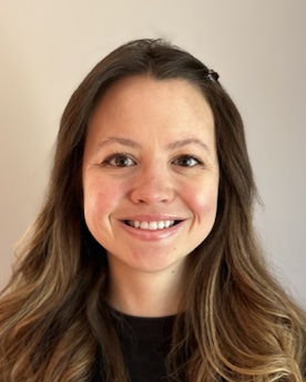
Awarded Organization: Massachusetts General Hospital
Awarded Department: Department of Molecular Biology
Sponsor: Dr. Luke ChaoAward Year Start: 2023
Current Title: Postdoctoral Fellow
Project Title: In situ cryoET analysis of cristae biogenesis and morphology
Mitochondria generate energy needed to power cells and multicellular organisms. Wrinkles in the inner mitochondrial membrane, known as cristae, concentrate molecular motors for energy production. However, it is unclear how the wrinkly cristae are formed. Dr. Michelle Fry will use a clever approach to investigate cristae formation in cells. She will introduce candidate protein/protein complexes into parasitic protist mitochondria. These mitochondria are smooth, making them amenable for testing with proteins are sufficient to generate cristae. Dr. Fry will use advanced electron microscopy techniques to image changes in mitochondrial morphology. Fry will conduct these studies in Dr. Luke Chao’s lab at Massachusetts General Hospital. These experiments will provide fundamental insights into mitochondrial biology and may provide clues for mitochondrial pathological dysfunction.
As a graduate student in Dr. Bil Clemons lab at the California Institute of Technology, Fry used structural biology to study the targeting of membrane proteins to the endoplasmic reticulum. Specifically, Dr. Fry captured several structural conformations of a protein chaperone, Get3. Fry demonstrated how conformational flexibility is important for Get3 to integrate multiple regulatory signals (binding partners, client proteins, nucleotide binding and hydrolysis). Dr Fry is now excited to use cryo-electron tomography to capture the conformational landscape of proteins that regulate mitochondrial cristae formation in cells.
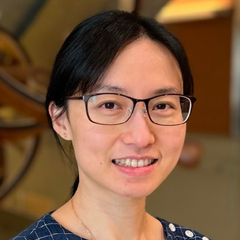
Awarded Organization: University of Washington
Awarded Department: Department of Genome Science
Sponsor: Dr. Jay ShendureAward Year Start: 2024
Current Title: Postdoctoral Fellow
Project Title: Deciphering the dynamic regulation of mitochondrial genomes
Mitochondria are cellular organelles that house their own DNA. There are hundreds to thousands of copies of the mitochondrial genome (mtDNA) in each cell. Often, mtDNA copies are not the same; rather, a fraction of them carries mutations. Moreover, the composition of mtDNA varies drastically across cells and cell types. Mitochondrial diseases manifest when the pathogenic mutations reach a high percentage in a substantial fraction of cells. However, it is still unclear how mtDNA mutations expand and how cell-to-cell variation of mtDNA composition is formed.
Dr. Yi Fu will address these questions in Dr. Jay Shendure’s lab at the University of Washington. Dr. Fu will develop a method to accurately genotype mtDNA at single-cell resolution and employ this method to monitor mtDNA mutations during differentiation. Fu will also combine this approach with CRISPR perturbation to identify factors that impact the mitochondrial mutation burden in various cell types. These experiments will uncover cell type-specific regulation of mitochondrial genome maintenance. Furthermore, Fu’s research may provide insight into novel therapeutic approaches for mtDNA-associated diseases.
Fu’s expertise in mtDNA stems from her graduate studies in Dr. Agnel Sfeir’s lab at New York University and Memorial Sloan Kettering Cancer Center. There Fu discovered that double-strand breaks in mtDNA activate the integrated stress response, highlighting the cellular program to cope with defective mitochondrial genome. Fu also investigated mtDNA deletions and their impact on cellular metabolism. Now, Fu will leverage genomics and single-cell technologies to elucidate the dynamic regulation of mtDNA during her postdoctoral research.